×

National Heart Foundation of Australia and Cardiac Society of Australia and New Zealand: Australian clinical guidelines for the management of acute coronary syndromes 2016

These clinical guidelines have been developed to assist in managing patients presenting with chest pain suspected to be caused by an acute coronary syndrome (ACS) and those with confirmed ACS. The development of these guidelines has been informed by reviews of the literature dealing with key aspects of chest pain assessment and ACS care, as well as broad consultation with local opinion leaders, stakeholder groups and the public. The recommendations focus on the core clinical and system-based components of care most associated with improved clinical outcomes. As such, these guidelines should be read in conjunction with the Acute coronary syndromes clinical care standard, developed by the Australian Commission on Safety and Quality in Health Care,1 and the Australian acute coronary syndromes capability framework, developed by the National Heart Foundation of Australia (NHFA).2 Guidance regarding both the strength of evidence supporting the recommendations and their potential impact on outcomes is provided to assist in informing clinical practice.3,4 Additional guidance regarding the timing and considerations informing the use of therapies and management strategies is given in the accompanying practice advice. A full version of the NHFA and Cardiac Society of Australia and New Zealand (CSANZ) Australian clinical guidelines for the management of acute coronary syndromes 2016 is available at: http://heartfoundation.org.au/for-professionals/clinical-information/acute-coronary-syndromes.

Methods

The NHFA, in partnership with the CSANZ, has undertaken an update to the NHFA/CSANZ Guidelines for the management of acute coronary syndromes 2006 and addenda of 2007 and 2011.57 The updated guideline will provide a synthesis of current evidence-based guidance for health professionals caring for patients with ACS.

The ACS Guideline Development Working Group comprised an Executive and the four writing groups of which it had oversight, covering the topics of chest pain, ST segment elevation myocardial infarction (STEMI), non-ST segment elevation ACS (NSTEACS) and secondary prevention. In addition, a Reference Group included representatives from stakeholder groups, potential endorsing organisations and regional experts. The Working Group comprised a broad mix of health professionals, including a general practitioner, general physician, cardiac surgeon, consumer representative, pathologist, ambulance service representative, cardiologists, emergency physicians, exercise physiologists and cardiac nurses.

The Working Group consulted state-based cardiac clinical networks and the Reference Group on the scope determination for the updated guideline. Based on this consultation, the expert Working Group generated clinical questions to inform the literature search of evidence required for the guideline’s development. The separate writing groups reviewed and graded the evidence, generated and graded recommendations, and produced draft sections for the four topic areas. The Executive group provided oversight for this process and approved the final document.

A draft of the guideline was open for a 30-day period of public consultation in April 2016 to capture stakeholder views and aid engagement with the guideline once completed. Attention has been paid to ensuring appropriate governance processes were in place, to ensure transparency, minimise bias, manage conflict of interest and limit other influences during guideline development.

Key evidence-based recommendations

Each recommendation is presented with a Grading of Recommendations Assessment, Development and Evaluation (GRADE) strength of recommendation (Appendix 1) and a National Health and Medical Research Council level of evidence (Level) (Appendix 2). Practice points (PPs) are also provided.

Assessment of possible cardiac causes of chest pain

  • It is recommended that a patient with acute chest pain or other symptoms suggestive of an ACS receives a 12-lead electrocardiogram (ECG), and this ECG is assessed for signs of myocardial ischaemia by an ECG-experienced clinician within 10 minutes of first acute clinical contact.8 GRADE: Strong; Level: IIIC

    • PP: Oxygen supplementation. The routine use of oxygen therapy among patients with a blood oxygen saturation (SaO2) level > 93% is not recommended, but its use when the SaO2 is below this level is advocated, despite the absence of clinical data.9,10 However, care should be exercised in patients with chronic obstructive pulmonary disease where the target SaO2 level is to be 88–92%.

    • PP: Initial aspirin therapy. In all patients with possible ACS and without contraindications, aspirin (300 mg orally, dissolved or chewed) should be given as soon as possible after presentation. Additional antiplatelet and anticoagulation therapy, or other therapies such as β-blockers, should not be given to patients without a confirmed or probable diagnosis of ACS.

  • A patient presenting with acute chest pain or other symptoms suggestive of an ACS should receive care guided by an evidence-based Suspected ACS Assessment Protocol (Suspected ACS-AP) that includes formal risk stratification.11 GRADE: Strong; Level: IA

    • PP: Selecting and implementing a Suspected ACS-AP. For hospitals using sensitive or highly sensitive troponin assays, the ADAPT or modified ADAPT protocol, respectively, identifies low risk patients (< 1% major adverse cardiac events [MACE] at 30 days) on the basis of negative troponin test results at both 0 and 2 hours, a Thrombolysis in Myocardial Infarction (TIMI) risk score of 0 (ADAPT) or 0 or 1 (modified ADAPT), and no ischaemic changes on ECG at both 0 and 2 hours.12,13

  • Using serial sampling, cardiac-specific troponin levels should be measured at hospital presentation and at clearly defined periods after presentation using a validated Suspected ACS-AP in patients with symptoms of possible ACS.14 GRADE: Strong; Level: IA

    • PP: Timing of troponin testing. Most patients with an underlying diagnosis of acute myocardial infarction (AMI) have elevated troponin levels within 3–6 hours of symptom onset, although some assays may not show elevated levels for up to 12 hours (Box 1). Validated rapid rule-in and rule-out algorithms for AMI incorporated into Suspected ACS-APs and/or using highly sensitive troponin assays may reduce the serial testing time to 1–2 hours after presentation.18,19,21,24,25 Incorporating sensitive or highly sensitive troponin assay results into the ADAPT or modified ADAPT protocol, respectively, allows early (2 hours after emergency department presentation) risk stratification.12,13

  • Non-invasive objective testing is recommended in intermediate risk patients, as defined by a validated Suspected ACS-AP, with normal serial troponin and ECG testing and who remain symptom free.26 GRADE: Weak; Level: IA

    • PP: Timing of testing. High risk patients require further objective testing during the index admission (Box 2). Intermediate risk patients may be safely accelerated for early inpatient testing or discharged for outpatient testing, ideally within 7 days but acceptable up to 14 days after presentation. Investigation before discharge from the emergency department is desirable among patients with characteristics associated with significant failure to re-attend for medical review, given the higher rates of MACE in such patients.27

  • Patients in whom no further objective testing for coronary artery disease is recommended are those at low risk, as defined by a validated Suspected ACS-AP: age < 40 years, symptoms atypical for angina, in the absence of known coronary artery disease, with normal troponin and ECG testing and who remain symptom free.26 GRADE: Weak; Level: III-3C

Diagnostic issues, risk stratification and acute management of ACS

  • The routine use of validated risk stratification tools for ischaemic and bleeding events (eg, GRACE score for ischaemic risk and CRUSADE score for bleeding risk) may assist in patient-centric clinical decision making in regards to ACS care.2830 GRADE: Weak; Level: IIIB

    • PP: Choice of risk score. For ischaemic risk, the GRACE risk score is superior to the TIMI risk score in terms of discriminating between high risk and intermediate or low risk patients.28 However, estimating risk of death or recurrent myocardial infarction (MI) for an individual patient depends on local validation. For bleeding risk, the CRUSADE risk score is preferred, although it has limited validation in the Australian setting.31

Acute reperfusion and invasive management strategies for ACS

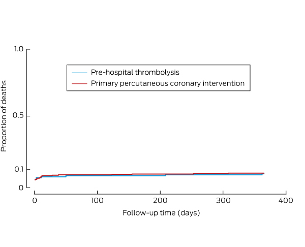
  • For patients with STEMI presenting within 12 hours of symptom onset, and in the absence of advanced age, frailty and comorbidities that influence the individual’s overall survival, emergency reperfusion therapy with either primary percutaneous coronary intervention (PCI) or fibrinolytic therapy is recommended.32,33 GRADE: Strong; Level: IA

    • PP: ECG interpretation. In situations where expertise in ECG interpretation may not be available, an electronic algorithm for ECG interpretation (coupled with review by an expert) can assist in diagnosing STEMI. Local or state care pathways should incorporate means for allowing expert ECG reading within 10 minutes of first contact, integrated with clinical decision making to enable timely reperfusion.

  • Primary PCI is preferred for reperfusion therapy in patients with STEMI if it can be performed within 90 minutes of first medical contact; otherwise, fibrinolytic therapy is preferred for those without contraindications.3436 GRADE: Strong; Level: IA

    • PP: Strategies for reducing the time to reperfusion therapy. Coordinated protocols with planned decision making that incorporates ambulance services and paramedics, first-responder primary care physicians, and emergency and cardiology departments are critical for achieving acceptable reperfusion times. Strategies need to be tailored to the local community and the distribution of emergency services. Strategies that effectively shorten the time to reperfusion include: developing hospital networks with pre-determined management pathways for reperfusion; pre-hospital ECG and single call catheter laboratory activation; pre-hospital fibrinolytic therapy administered by suitably trained clinicians (eg, paramedics); the bypassing, where appropriate, of non-PCI-capable hospitals; and bypassing the emergency department on arrival in PCI-capable centres. Furthermore, an established capability for timely expert consultation for complex clinical scenarios is highly desirable. In the context of a system-based approach to reperfusion, the capacity for continuous audit and feedback is also advocated.

  • Among patients treated with fibrinolytic therapy who are not in a PCI-capable hospital, early or immediate transfer to a PCI-capable hospital for angiography, and PCI if indicated, within 24 hours is recommended.37 GRADE: Weak; Level: IIA

  • Among patients treated with fibrinolytic therapy, for those with ≤ 50% ST recovery at 60–90 minutes and/or with haemodynamic instability, immediate transfer for angiography with a view to rescue angioplasty is recommended.38 GRADE: Strong; Level: IB

    • PP: Hospital networks. Systems of care should be developed to provide advice and enable, when appropriate, immediate or early transfer for angiography of patients treated with fibrinolytic therapy who are not in a PCI-capable hospital.

  • Among high and very high risk patients with NSTEACS (except type 2 MI [secondary to ischaemia due to either increased oxygen demand or decreased supply]), a strategy of angiography with coronary revascularisation (PCI or coronary artery bypass grafting [CABG]), where appropriate, is recommended.39 GRADE: Strong; Level: IA

    • PP: Mode of revascularisation. Patient comorbidities, fitness for major surgery and coronary anatomy are the main determinants. Urgent revascularisation with CABG may be indicated for patients with failed PCI, cardiogenic shock or mechanical defects resulting from MI (eg, septal, papillary muscle or free-wall rupture). A combined Heart Team approach may provide the best consensus decision about the care of an individual patient.

    • PP: Invasive management for type 2 MI. Type 2 MI remains a challenging diagnosis, and no trials have examined the benefits of a routine invasive strategy in patients with type 2 MI. In the absence of any trial evidence, angiography with a view to revascularisation may be considered if there is ongoing ischaemia or haemodynamic compromise despite adequate treatment of the underlying acute medical problem that provoked the type 2 MI.

  • Patients with NSTEACS who have no recurrent symptoms and no risk criteria are considered at low risk of ischaemic events and can be managed with a selective invasive strategy guided by provocative testing for inducible ischaemia.39 GRADE: Strong; Level: IA

  • Very high risk patients: Among patients with NSTEACS with very high risk criteria (ongoing ischaemia, haemodynamic compromise, arrhythmias, mechanical complications of MI, acute heart failure, recurrent dynamic or widespread ST segment and/or T wave changes on ECG; Box 3), an immediate invasive strategy is recommended (ie, within 2 hours of admission).40 GRADE: Strong; Level: IIC

  • High risk patients: In the absence of very high risk criteria, for patients with NSTEACS with high risk criteria (GRACE score > 140, dynamic ST segment and/or T wave changes on ECG or rise and/or fall in troponin compatible with MI; Box 3), an early invasive strategy is recommended (ie, within 24 hours of admission).40 GRADE: Weak; Level: IC

  • Intermediate risk patients: In the absence of high risk criteria, for patients with NSTEACS with intermediate risk criteria (such as recurrent symptoms or substantial inducible ischaemia on provocative testing; Box 3), an invasive strategy is recommended (ie, within 72 hours of admission).4042 GRADE: Weak; Level: IIC

Pharmacology for ACS

  • Aspirin 300 mg orally (dissolved or chewed) initially, followed by 100–150 mg/day, is recommended for all patients with ACS, in the absence of hypersensitivity.43 GRADE: Strong; Level: IA

  • Among patients with confirmed ACS at intermediate to very high risk of recurrent ischaemic events, use of a P2Y12 inhibitor (ticagrelor 180 mg orally, then 90 mg twice a day; or prasugrel 60 mg orally, then 10 mg daily; or clopidogrel 300–600 mg orally, then 75 mg daily) is recommended in addition to aspirin (ticagrelor or prasugrel preferred; see below).4447 GRADE: Strong; Level: IA

    • PP: Choosing between P2Y12 inhibitors. Given their superior efficacy, ticagrelor and prasugrel are the preferred first-line P2Y12 inhibitors. Use of ticagrelor is advised for a broad spectrum of patients with STEMI or NSTEACS who are at intermediate to high risk of an ischaemic event, in the absence of atrioventricular conduction disorders (second and third degree atrioventricular block) and asthma or chronic obstructive pulmonary disease. Prasugrel may be considered for patients who have not received a P2Y12 antagonist and in whom PCI is planned, but it should not be used for patients > 75 years of age, of low bodyweight (< 60 kg) or with a history of transient ischaemic attack or stroke. Use of either prasugrel or ticagrelor, rather than clopidogrel, is also recommended for patients who have experienced recurrent events while taking clopidogrel or who have experienced stent thrombosis. Clopidogrel is recommended for patients who cannot receive ticagrelor or prasugrel, as an adjunctive agent with fibrinolytic therapy or for those requiring oral anticoagulation (refer to relevant prescribing information documentation). Ticagrelor or clopidogrel should be commenced soon after diagnosis, but due consideration should be given to ischaemic and bleeding risks, the likelihood of need for CABG (more likely in patients with extensive ECG changes, ongoing ischaemia or haemodynamic instability) and the delay to angiography. Prasugrel should be commenced immediately after diagnosis among patients undergoing primary PCI for STEMI, or after the coronary anatomy is known among those undergoing urgent PCI. Initiation of prasugrel before coronary angiography outside the context of primary PCI is not recommended.

    • PP: Combination of P2Y12 inhibition with long term anticoagulation. Among patients with an indication for oral anticoagulation, a careful assessment of thrombotic and bleeding risks is required, using CHA2DS2-VASc and HAS-BLED scores, respectively. The following advice is based on consensus opinion. In patients with a strong indication for long term anticoagulation (ie, mechanical heart valves, atrial fibrillation with CHA2DS2-VASc score ≥ 2), the anticoagulant should be continued at a reduced dose, and clopidogrel, rather than ticagrelor or prasugrel, should be used for these patients. The duration of triple therapy (ie, aspirin, clopidogrel and oral anticoagulation) should be determined by the bleeding risk.

  • Intravenous glycoprotein IIb/IIIa inhibition in combination with heparin is recommended at the time of PCI among patients with high risk clinical and angiographic characteristics or for treating thrombotic complications among patients with ACS.48 GRADE: Strong; Level: IB

  • Either unfractionated heparin or enoxaparin is recommended in patients with ACS at intermediate to high risk of ischaemic events.49,50 GRADE: Strong; Level: IA

    • PP: Choosing between indirect thrombin inhibitors. Enoxaparin may be preferred over unfractionated heparin as it does not require monitoring of partial thromboplastin time and is simpler to administer. Swapping between enoxaparin and unfractionated heparin has been shown to increase bleeding risk and is not recommended.

  • Bivalirudin (0.75 mg/kg intravenously with 1.75 mg/kg/h infusion) may be considered as an alternative to glycoprotein IIb/IIIa inhibition and heparin among patients with ACS undergoing PCI with clinical features associated with an increased risk of bleeding events.51 GRADE: Weak; Level: IIB

Discharge management and secondary prevention

  • Aspirin (100–150 mg/day) should be continued indefinitely unless it is not tolerated or an indication for anticoagulation becomes apparent.43 GRADE: Strong; Level: IA

  • Clopidogrel should be prescribed if aspirin is contraindicated or not tolerated. GRADE: Strong; Level: IA

  • Dual antiplatelet therapy with aspirin and a P2Y12 inhibitor (clopidogrel or ticagrelor) should be prescribed for up to 12 months in patients with ACS, regardless of whether coronary revascularisation was performed. The use of prasugrel for up to 12 months should be confined to patients receiving PCI. GRADE: Strong; Level: IA

  • Consider continuation of dual antiplatelet therapy beyond 12 months if ischaemic risks outweigh the bleeding risk of P2Y12 inhibitor therapy; conversely, consider discontinuation if bleeding risk outweighs ischaemic risks.52 GRADE: Weak; Level: IIC

  • Initiate and continue indefinitely, the highest tolerated dose of an HMG-CoA (3-hydroxy-3-methylglutaryl-coenzyme A) reductase inhibitor (statin) for a patient following hospitalisation with ACS, unless contraindicated or there is a history of intolerance.53 GRADE: Strong; Level: IA

    • PP: Target cholesterol levels. There is additional benefit from progressive lowering of cholesterol levels, with no apparent lower limit. Within the context of an individualised care plan, a target low density lipoprotein cholesterol level of less than 1.8 mmol/L is suggested in the first instance.

  • Initiate treatment with vasodilatory β-blockers in patients with reduced left ventricular systolic function (left ventricular ejection fraction ≤ 40%) unless contraindicated.54 GRADE: Strong; Level: IIA

  • Initiate and continue angiotensin-converting enzyme inhibitors (or angiotensin receptor blockers) in patients with evidence of heart failure, left ventricular systolic dysfunction, diabetes, anterior MI or co-existent hypertension.55 GRADE: Strong; Level: IA

  • Attendance at cardiac rehabilitation or undertaking a structured secondary prevention service is recommended for all patients hospitalised with ACS.56,57 GRADE: Strong; Level: IA

    • PP: Individualisation of cardiac rehabilitation or secondary prevention service referral. A wide variety of prevention programs improve health outcomes in patients with coronary disease. After discharge from hospital, patients with ACS and, where appropriate, their companion(s) should be referred to an individualised preventive intervention according to their personal preference and values and the available resources. Services can be based in the hospital, primary care, the local community or the home.

System considerations

  • Continuous audit and feedback systems, integrated with work routines and patient flows, are strongly advocated to support quality assurance initiatives and provide data confirming continued, cost-efficient improvement in patient outcomes as a result of new innovations in care.

Box 1 –
Timing of troponin testing

Timing of sampling

Strategy*

Assays


0 hour (single sample)

Patients whose pain and symptoms resolved 12 hours prior to testing (cut points are the assay-specific 99th percentile

Both sensitive and highly sensitive (HS) assays

0 hour (single sample)

Patients with value < LoD of the specific assay (not > 99th percentile cut point) and symptom onset > 3 hours1517

HS assays

0 hour and 1 hour after presentation

Rule-in and rule-out AMI algorithms (cut points are assay-specific and not the 99th percentile)1820

HS assays

0 and 2 hours after presentation

ADAPT protocol13

Sensitive assays

Modified ADAPT protocol12,21(cut points are the assay-specific 99th percentile)

HS assays

0 and ≥ 3 hours after presentation

Previous NHFA protocol7

HS assays

HEART Pathway22,23 (cut points are the assay-specific 99th percentile)

Both sensitive and HS assays

0 and ≥ 6–12 hours after presentation

Rule-in and rule-out AMI algorithms5 (cut points are the assay-specific 99th percentile)

Sensitive and point-of-care assays


ADAPT = 2-Hour Accelerated Diagnostic Protocol to Assess Patients with Chest Pain Symptoms Using Contemporary Troponins as the Only Biomarker. AMI = acute myocardial infarction. HEART = History, Electrocardiogram, Age, Risk factors and Troponin. LoD = limit of detection. NHFA = National Heart Foundation of Australia. * With concurrent clinical risk stratification. † Reports on the use and outcomes of the biomarker strategy in clinical practice are not currently available.

Box 2 –
Risk classification for possible cardiac causes of chest pain


High risk

  • Ongoing or recurrent chest discomfort despite initial treatment
  • Elevated cardiac troponin level
  • New ischaemic changes on electrocardiogram (ECG), such as persistent or dynamic ECG changes of ST segment depression ≥ 0.5 mm; transient ST segment elevation (≥ 0.5 mm) or new T wave inversion ≥ 2 mm in more than two contiguous leads; or ECG criteria consistent with Wellens syndrome
  • Diaphoresis
  • Haemodynamic compromise — systolic blood pressure < 90 mmHg, cool peripheries, Killip Class > I and/or new onset mitral regurgitation
  • Sustained ventricular tachycardia
  • Syncope
  • Known left ventricular systolic dysfunction (left ventricular ejection fraction ≤ 40%)
  • Prior acute myocardial infarction, percutaneous coronary intervention or coronary artery bypass grafting

Low risk

  • Age < 40 years
  • Symptoms atypical for angina
  • Remain symptom free
  • Absence of known coronary artery disease
  • Normal troponin level
  • Normal ECG

Intermediate risk

  • Neither high risk nor low risk criteria

Box 3 –
Markers of increased risk of mortality and recurrent events among patients with confirmed acute coronary syndrome

Risk classification

Clinical characteristic


Very high

  • Haemodynamic instability, heart failure, cardiogenic shock or mechanical complications of myocardial infarction (MI)
  • Life-threatening arrhythmias or cardiac arrest
  • Recurrent or ongoing ischaemia (ie, chest pain refractory to medical treatment) or recurrent dynamic ST segment and/or T wave changes, particularly with intermittent ST segment elevation, de Winter T wave changes or Wellens syndrome, or widespread ST segment elevation in two coronary territories

High

  • Rise and/or fall in troponin level consistent with MI
  • Dynamic ST segment and/or T wave changes with or without symptoms
  • GRACE score > 140

Intermediate

  • Diabetes mellitus
  • Renal insufficiency (glomerular filtration rate < 60 mL/min/1.73 m2)
  • Left ventricular ejection fraction ≤ 40%
  • Prior revascularisation: percutaneous coronary intervention or coronary artery bypass grafting
  • GRACE score > 109 and < 140

GRACE = Global Registry of Acute Coronary Events.

The uptake of coronary fractional flow reserve in Australia in the past decade

The use of coronary pressure wires (or fractional flow reserve [FFR]) has been shown to reduce the frequency of major adverse cardiac events and of unnecessary stent procedures, and to lower treatment costs in both the public and private sectors in Australia.13 FFR is a tool for assessing physiological ischaemia in coronary artery stenosis, measuring pre- and post-stenosis pressures during adenosine-induced hyperaemia. Because it is evidence-based and quantifiable, it may be discussed during the upcoming Medicare reform. Data on its uptake across Australia, however, have not been published.

We examined trends in FFR use after its addition to the Medicare Benefits Schedule 10 years ago. We analysed Australian Government Department of Human Services data on Medicare items for coronary flow reserve, coronary angiography and percutaneous coronary angiography.

A total of 14 160 FFR services were processed by Medicare during the past 10 years. FFR use grew during this period, with a mean annual increase of 55%, from 131 services in 2007 to 3869 in 2015 (non-parametric analysis, P = 0.004). Time series analysis identified a Gompertz non-linear trend of FFR against time, indicating that national FFR use is continuing to increase, although growth began to slow in 2014. Further, FFR use increased on a population basis by an average of 45% each year, from 1 per 100 000 in 2007 to 16 per 100 000 in 2015, when these figures were highest in New South Wales (23 per 100 000) and Queensland (19 per 100 000) (Box).

The national rate of FFR per coronary angiogram increased from 0.02% in 2006 to 4.8% in 2015 (P = 0.004), when the highest rate was in NSW (5.8%). The rate of FFR per percutaneous coronary intervention (PCI) increased from 0.1% in 2006 to 19.2% in 2015 (P = 0.004), when the highest rate was in Queensland (26.6%). In 2015, there were 5.2 PCIs per FFR used; the rate was not related to the population size of the state or territory (Spearman non-parametric correlation, ρS = 0.07; P = 0.87) or to total PCI use (ρS = 0.12, P = 0.78). There was marked variation between states and territories (Box), highlighting heterogeneity across Australia in the use of FFR.

The data summarised in the Box allow operators and hospitals to compare their use of FFR with state and national averages, and they facilitate more standardised care across Australia. Barriers to the uptake of FFR include operator and centre experience, availability and cost. It has been suggested that FFR is discouraged by the lower remuneration received if stenting is not performed.4 From a national perspective, however, there is a mean saving of $1200 per patient in the public sector and $5000 per patient in the private sector when FFR makes stenting unnecessary,2 representing a total annual saving of $4 million.4

The use of FFR across Australia is heterogeneous, but it has grown over the past decade, both in absolute numbers and as proportions of coronary angiograms and PCI.

Box –
Summary of Medicare items for use of a coronary pressure wire (fractional flow reserve [FFR]) processed during 2015

Australia

NSW

Vic

Qld

SA

WA

Tas

ACT

NT


Total FFR services

3869

1764

688

944

146

225

77

11

14

FFR per 100 000 population

16

23

11

19

8

9

15

3

6

FFR per angiogram

1/21 (4.8%)

1/17 (5.8%)

1/28 (3.6%)

1/17 (5.7%)

1/30 (3.4%)

1/29 (3.5%)

1/21 (4.8%)

1/83 (1.2%)

1/38 (2.7%)

FFR per percutaneous coronary intervention

1/5.2 (19.2%)

1/4.6 (21.6%)

1/7.0 (14.3%)

1/3.8 (26.6%)

1/6.8 (14.7%)

1/7.7 (13.0%)

1/6.2 (16.2%)

1/34.1 (2.9%)

1/4.3 (23.3%)


Source: Australian Government Department of Human Services. Medicare Australia Statistics, medical item reports (http://medicarestatistics.humanservices.gov.au/statistics/mbs_item.jsp) for coronary flow reserve (item number, 38241), coronary angiogram (38215, 38218, 38220, 38222, 38225, 38228, 38231, 38234, 38237, 38240, 38246) and percutaneous coronary angiogram (38243, 38246).

Pre-hospital thrombolysis in ST-segment elevation myocardial infarction: a regional Australian experience

The known Pre-hospital thrombolysis in patients with ST-segment elevation myocardial infarction (STEMI) has been found to be an effective strategy in randomised controlled trials.

The new Pre-hospital thrombolysis is safe and effective in the real world setting, especially in a region where transport distances to the cardiac catheterisation laboratory are great.

The implications Pre-hospital thrombolysis can be implemented in regions where timely primary percutaneous coronary intervention for STEMI patients is not available.

Primary percutaneous coronary intervention (PCI), if performed in a timely manner, is the preferred reperfusion strategy for patients with an ST-segment elevation myocardial infarction (STEMI).1 However, physicians in regions geographically isolated from a primary PCI centre are unable to perform PCI rapidly, and the optimal reperfusion strategy is therefore undefined. Recent guidelines have highlighted the importance of systems-based approaches to STEMI management, taking into account regional characteristics.2 In Australia, many patients are a long way from a primary PCI centre, and these patients usually receive thrombolysis in hospitals that are not PCI-capable. However, if reperfusion with thrombolysis fails or a contraindication prevents its application, there can be long delays in reaching a cardiac catheterisation laboratory (CCL) for PCI.3 Further, a pharmaco-invasive (PI) strategy, including routine cardiac catheterisation within 24 hours of thrombolysis, was found to achieve better outcomes than ischemia-driven angiography.4 This strategy is especially relevant in areas where geographic access and logistical delays that increase the time to treatment (eg, rural areas, large urban areas with traffic congestion) may reduce some of the benefits of primary PCI in clinical practice.5

The international Strategic Reperfusion Early After Myocardial Infarction (STREAM) trial, in which STEMI patients who were unable to undergo primary PCI within one hour of presentation were randomised to either primary PCI or a PI strategy, confirmed that outcomes of the PI strategy at one year were not inferior to those of primary PCI.6,7 However, the STREAM trial did not include all patients who presented with STEMI; although most of those randomised underwent randomisation in the ambulance setting (81%), the patients included in the trial had presented within 3 hours of symptom onset (compared with 6–12 hours for the STEMI patients who were not included), and the median time from presentation to PCI in the PI arm was 10 hours, which may not be achievable in a real world setting. Similarly, geographical and logistical constraints that are important factors in different parts of the world, such as distance and regional resources, were not considered in the STREAM trial.

The case series described in this article was from the Hunter New England Local Health District (HNELHD), which has a complex geography and limited resources. The area it covers (130 000 km2) is comparable in size with England, but it has only one continuously operating CCL; it is located in one corner of the health district (in Newcastle), and patients with a STEMI may be as far as 600 km away (Box 1). In 2008, we implemented a system of care in HNELHD for providing rapid reperfusion to STEMI patients throughout this large district. Modelled on the Emergency Triage of Acute Myocardial Infarction study in the Northern Sydney Area Health Service, which achieved reduced delays and improved outcomes for patients with primary angioplasty,9 our system involved pre-hospital diagnosis and triage by paramedic staff, followed by their allocating the patients to either PHT or primary PCI according to their travel time to the CCL in our large regional health district. The analysis reported in this article compares the safety and effectiveness of pre-hospital assessment and allocation to pre-hospital thrombolysis (PHT) or primary angioplasty by paramedics.10

Methods

Study design

Hunter AMI was a prospective, non-randomised, consecutive, single-centre case series of STEMI patients who had been diagnosed on the basis of a pre-hospital ECG performed by paramedics. Data were collected electronically through the collaboration of the New South Wales Ambulance and the HNELHD, and data were independently analysed by the Clinical Research Design, IT and Statistical Support (CReDITSS) unit at the Hunter Medical Research Institute.

Study patients

For all patients with symptoms or signs suggesting myocardial infarction, paramedics recorded a 12-lead electrocardiogram (ECG; LIFEPAK 15 monitor system, Physio-Control Australia). Posterior and right-sided leads were not recorded by the paramedics for infero-posterior or right ventricular myocardial infarction. If the ECG indicated a possible STEMI according to the Glasgow algorithm,11,12 it was transmitted to the on-call cardiologist or cardiology advanced trainee at the John Hunter Hospital in Newcastle; the doctor and the paramedic then discussed the case, and reperfusion of the patient started. For patients who could reach a CCL within 60 minutes of first medical contact (FMC) or for whom there was any contraindication to thrombolysis (irrespective of FMC-to-CCL door time), reperfusion therapy consisted of primary PCI at the John Hunter Hospital. PHT (fibrinolysis with tenecteplase administered by paramedics) was administered by paramedics to patients for whom the FMC-to-CCL door time exceeded 60 minutes, and was followed by transfer to the PCI-capable centre (Appendix 1). All consecutive STEMI patients who had been diagnosed by pre-hospital ECG were included in our analysis.

FMC was defined as the first face-to-face contact made by paramedics with the patient. Total ischaemic time was defined as the time from symptom onset to balloon inflation for the primary PCI group, and to injection of tenecteplase for the PHT group. Hypertension was diagnosed if blood pressure greater than 140/90 mmHg was measured on two or more occasions in the hospital, or if the patient was already being treated with antihypertensive medication.13,14 Diabetes mellitus was diagnosed if two of the following applied: symptoms of hyperglycaemia; fasting sugar level > 7 mmol/L; 2-hour post-prandial sugar level > 11.1 mmol/L; HbA1c level > 6.5 mmol/mol.15 Cardiogenic shock was defined as persistent hypotension (systolic blood pressure < 90 mmHg or mean arterial pressure 30 mmHg lower than baseline).16 Left ventricular ejection fraction was recorded during the index hospitalisation by transthoracic echocardiography, using Simpson’s biplane method.

Study therapies

We present the outcomes of PHT and primary PCI performed according to our local guideline-based protocol, which includes concomitant dual antiplatelet and anticoagulant therapy. Adjunct administration of a glycoprotein IIb/IIIa antagonist was at the discretion of the intervening cardiologist. In the PHT arm, tenecteplase was administered at a weight-based dose (55 to < 60 kg, 30 mg; 60 to < 70 kg, 35 mg; 70 to < 80 kg, 40 mg; 80 to < 90 kg, 45 mg; ≥ 90 kg, 50 mg). The tenecteplase dose administered to patients aged 75 years or more has since been reduced by half. Anticoagulant therapy consisted of a 30 mg intravenous bolus of enoxaparin (omitted for patients aged 75 years or more) followed by subcutaneous injection of 1 mg/kg bodyweight (0.75 mg/kg for patients aged 75 years or more) twice each day. Antiplatelet therapy consisted of a 300 mg loading dose of clopidogrel (omitted for patients aged 75 years or more) followed by 75 mg daily, together with an initial 300 mg aspirin dose, immediately followed by 100 mg daily. Patients in the PHT arm with electrical or haemodynamic instability, ongoing ischaemic symptoms, or less than 50% ST-segment resolution within 90 minutes of thrombolysis underwent rescue PCI.

Primary endpoints

The primary efficacy endpoint was 12-month all-cause mortality. The primary safety endpoint was bleeding, categorised according to Thrombolysis in Myocardial Infarction (TIMI) study group criteria.17

Statistical analysis

Summary statistics for patient characteristics in each of the two groups are presented. Normality of distribution was assessed with the Shapiro–Wilk test. Between-group comparisons of continuous variables used independent sample t tests for parametric data and pairwise comparison median tests for non-parametric data. The Pearson χ2 test assessed differences between categorical variables. Nelson–Aalen survival distributions were estimated, and adjusted using Cox proportional hazards regression models. Predictors of 12-month mortality were assessed using logistic regression. All statistical analyses were conducted in Stata 14 (StataCorp) and SAS 9.4 (SAS Institute).

Ethics approval

The study protocol was approved by the Hunter New England Human Research Ethics Committee in August 2014 (reference, 14/06/18/5.09).

Results

From 1 August 2008 to 31 August 2013, 484 patients with STEMI diagnosed by pre-hospital ECG were allocated to PHT (150 patients) or primary PCI (334 patients) on the basis of estimated FMC-to-CCL door times or any contraindication to thrombolysis (Appendix 2). There were more patients in the primary PCI group with diabetes mellitus or hypertension, and fewer with hypercholesterolaemia (Box 2).

The median time from FMC to the start of reperfusion was 35 minutes (IQR, 28–43 min) for bolus tenecteplase and 130 minutes (IQR, 100–150 min) for balloon inflation (P = 0.001). The median time between symptom onset and treatment was correspondingly shorter in the PHT group (94 [IQR, 65–127] v 180 [IQR, 140–265] minutes) (Box 2). The median distance of patients in the PHT group from the CCL was 119 km (range, 8–483 km). The median time from symptom onset to angiography was thus longer in the PHT group than in the primary PCI group, with a delay of 4 hours for the 27% of patients who required rescue or urgent intervention, and of 49 hours for the other patients in this group. Significantly, more open vessels (TIMI flow grade of 2 or more) were found on first angiography in the PHT than in the primary PCI group (45% v 8%; P < 0.001) (Box 3).

There were three ischaemic strokes and two transient ischaemic attacks in the primary PCI group and none in the PHT group. Four of the five patients who suffered a transient ischaemic attack or stroke had atrial fibrillation, either before or detected during the admission. Bleeding was significantly more frequent in the PHT group than in the primary PCI group (9.3% v 5.1%; P = 0.001) with two (1.3%) TIMI major bleeds and one (0.7%) intracranial haemorrhage in the PHT group (Box 4).

Of the total cohort of 484 patients, 34 (7.0%) died within 12 months (the primary efficacy endpoint): 10 of 150 patients (6.7%) in the PHT group and 24 of 334 (7.2%) in the primary PCI group (relative risk in the PHT group, 0.93; 95% CI, 0.45–1.9; P = 0.84) (Box 5). Anterior STEMI, cardiogenic shock, and hypertension were independent predictors of mortality for the total cohort of 484 patients in the multivariate analysis (Box 6). Age, bleeding, ejection fraction and femoral access were significant only in the univariate analysis (data not shown).

Discussion

Our study produced several important findings. First, delivery of PHT by paramedics, based on algorithm assessment of 12-lead ECGs, is feasible and safe in regional Australia. Second, 12-month mortality for patients remote from a CCL who receive PHT and are then transferred to a PCI-capable centre is similar to that for patients near the CCL who receive primary PCI.

Our practice is to administer fibrinolysis to STEMI patients who are remote from the CCL if there is no contraindication, and to then assess clinical and ECG signs of reperfusion. If they were successfully reperfused, we did not mandate immediate cardiac catheterisation but instead brought them to the CCL as soon as convenient, provided they remained clinically stable in the meantime. Those for whom reperfusion was not successful or chest pain recurred were urgently transferred to the CCL for rescue PCI.

To place our findings in context, we compared them with the outcomes of the STREAM trial.7 Our PHT group had some notable differences from the STREAM PI group: our patients were older, if not statistically significantly (62 years [SD, 13] v 60 years [SD, 12]), and a greater proportion of our patients were aged 75 years or more (18% v 14%); we also had a higher proportion of patients with cardiogenic shock (5.3% v 0.1%). Sex balance, rates of diabetes, hypertension, and prior coronary artery bypass graft surgery, and symptom onset-to-treatment times were similar for our PHT patients and the STREAM fibrinolysis group. The median time to angiography for our PHT group was 28 hours, compared with 10 hours in the STREAM fibrinolysis arm. Despite these differences, which indicate a higher risk level for our patients, survival at 12 months in the two fibrinolysis groups was identical (93.3%).

All-cause mortality at 12 months was similar for both treatment groups in our study, but this was not a randomised controlled trial, so that conclusions about treatment efficacy should not be drawn. The safety of the pre-hospital assessment and treatment, on the other hand, is reassuring. As expected, there was a significantly higher bleeding complication rate in the PHT group, but only one intracranial haemorrhage. There was a higher stroke rate in the primary PCI group; most events occurred in association with atrial fibrillation, but this finding requires further follow-up assessment of potential contributors.

The optimal ECG algorithm for the detection of STEMI is unknown. For ease of use in the field, we chose to perform standard 12-lead ECGs without right ventricular or posterior leads. For their interpretation, we use the Glasgow algorithm because of its specificity.10 It remains unknown whether the addition of right ventricular or posterior leads, or the use of another algorithm, would improve the sensitivity or specificity of detection in this setting.

The transport times for the primary PCI group in our study were considerably longer than the 60-minute FMC-to-CCL goal. The decision to administer or withhold PHT was driven mainly by the estimated FMC-to-CCL time. In our protocol, the approximate time to the CCL is estimated by the paramedics, as their knowledge of prevailing weather and traffic conditions means they are in the best position to do so. Their decision is then discussed with the cardiologist or advanced trainee. There may be some bias towards underestimating transport times, but, more importantly, there are inherent difficulties in adhering to these guidelines. First, patients who had any contraindication to fibrinolysis underwent primary PCI, regardless of the estimated FMC-to-CCL time, and this would increase overall transport times for the primary PCI group. Second, clinical factors (eg, treatment of arrhythmias, resuscitation from cardiac arrest) sometimes interfere with transport, also prolonging transfer times. It is therefore important that actual transport times are reported, not just the goal transfer times. It is hoped that knowledge of both will help when devising systems of care for other Australian regions.

The long distances to the CCL for the patients in our PHT group underscore the importance of implementing systems of care in areas with complex geography that achieve optimal outcomes for STEMI patients. The median times from symptom onset to treatment in both our treatment groups were comparable with those of the STREAM groups. This finding suggests that total ischaemic time is the interval with the greatest prognostic impact, and that we should focus on reducing this period, not than just door-to-balloon times. While medical practitioners can influence treatment intervals after FMC, community education campaigns that encourage patients to call an ambulance as soon as they have chest pain are needed for reducing the time from symptom onset to FMC.

Our study had some important limitations. First, it was a non-randomised, consecutive case series study. We present the mortality associated with two therapies, but baseline and other differences between the two groups may have contributed to the different outcomes. Second, our study was underpowered for detecting small differences in mortality, so that our results should be interpreted with caution. It is nevertheless hoped that our data can inform systems of care for patients with STEMI throughout Australia.

In conclusion, our real world experience showed that PHT delivered by paramedics followed by early transfer to a PCI-capable centre is a safe and effective reperfusion strategy for patients remote from primary PCI centres.

Box 1 –
The Hunter New England Local Health District, about 600 km from north to south. The John Hunter Hospital is located in Newcastle, in the bottom right hand corner of the map*


* Adapted from: Eastwood et al (2010).8

Box 2 –
Baseline characteristics of the pre-hospital thrombolysis (PHT) and primary percutaneous coronary intervention (PCI) groups

All patients

PHT group

Primary PCI group

P


Total number

484

150

334

Age (years), mean (SD)

64 (13)

62 (13)

65 (13)

0.3

≥ 75 years

115 (24%)

27 (18%)

88 (26%)

0.05

Sex (males)

365 (75%)

114 (7%)

251 (75%)

0.8

Systolic blood pressure (mmHg), mean (SD)

130 (24)

125 (25)

131 (24)

0.1

Cardiogenic shock

30 (6.2%)

8 (5.3%)

22 (6.5%)

0.8

Anterior STEMI

188 (39%)

55 (37%)

133 (40%)

0.5

Cardiovascular history

Coronary artery disease

96 (20%)

28 (19%)

68 (20%)

0.9

Prior coronary artery bypass graft surgery

13 (2.7%)

2 (1.3%)

11 (3.3%)

0.2

Hypertension

267 (55%)

64 (43%)

203 (61%)

< 0.001

Diabetes mellitus

97 (20%)

20 (13%)

77 (23%)

0.01

Smoking

213 (44%)

67 (45%)

146 (44%)

0.8

Hypercholesterolaemia

198 (41%)

76 (51%)

122 (36%)

0.002

Body mass index (kg/m2), mean (SD)

29 (5)

28 (6)

29 (5)

0.08

First medical contact to treatment (min), median (IQR)

105 (49–140)

35 (28–43)

130 (100–150)

0.001

Symptom to treatment (min), median (IQR)

155 (107–235)

94 (65–127)

180 (140–265)

< 0.001

Symptom to first medical contact > 3 h

83 (17%)

14 (9.3%)

69 (21%)

0.002

Ejection fraction, mean (SD)

48% (8)(n = 332)

49% (10)(n = 236)

47% (7)(n = 96)

0.01

Peak troponin (ng/mL), mean (SD)

48 (32)

44 (28)

50 (35)

0.04

Haemoglobin (g/L), mean (SD)

144 (48)

145 (43)

143 (56)

0.2

Creatinine (μmol/L), mean (SD)

95 (31)

89 (20)

98 (34)

0.006

Length of stay (days), mean (SD)

4 (3)

4 (3)

4 (3)

1.0


STEMI = ST-segment elevation myocardial infarction.

Box 3 –
Angiographic characteristics of the pre-hospital thrombolysis (PHT) and primary percutaneous coronary intervention (PCI) groups

PHT group

Primary PCI group

P


Total number

150

334

Coronary angiography undertaken

138 (92%)

334 (100%)

< 0.001

Rescue PCI undertaken

37 (27%)

NA

Symptom onset to angiography (h), median (IQR)

28 (6–70)

3.5 (2.2–4.2)

< 0.001

Time to rescue PCI (h), median (IQR)

4 (3–5)

NA

Initial TIMI flow score ≥ 2

67 (45%)

27 (8.1%)

< 0.001

PCI/CABG undertaken

97 (65%)

307 (92%)

< 0.001

Femoral access

52 (38%)

153 (46%)

0.08

IIb/IIIa antagonist administered

7 (4.7%)

151 (45.2%)

0.004


CABG = coronary artery bypass graft surgery; NA = not applicable; TIMI = Thrombolysis in Myocardial Infarction study group.

Box 4 –
Primary efficacy and safety outcomes for the pre-hospital thrombolysis (PHT) and primary percutaneous coronary intervention (PCI) groups

PHT group

Primary PCI group

P


Primary efficacy outcome

12-month all-cause mortality

10 (6.7%)

24 (7.2%)

0.84

Primary safety outcomes

Intracranial haemorrhage

1 (0.7%)

0

Total bleeding (TIMI bleeding criteria)

14 (9.3%)

17 (5.1%)

0.001

TIMI major bleeding

2 (14%*)

0

TIMI minor bleeding

5 (36%*)

9 (53%*)

0.005

TIMI minimal bleeding

7 (50%*)

8 (47%*)

0.5


TIMI = Thrombolysis in Myocardial Infarction study group. * Percentage of all bleeding.

Box 5 –
Nelson–Aalen cumulative hazard estimates for patients receiving pre-hospital thrombolysis or primary percutaneous coronary intervention

Box 6 –
Independent predictors of mortality in 484 patients with ST-segment elevation myocardial infarction (multivariate analysis)

Odds ratio (95% CI)

P


Anterior STEMI

2.4 (1.2–4.8)

0.01

Cardiogenic shock

8.5 (3.4–21.3)

< 0.001

Hypertension

4.2 (1.8–10)

0.001


STEMI = ST-segment elevation myocardial infarction.

Variation in coronary angiography rates in Australia: correlations with socio-demographic, health service and disease burden indices

The known Angiography rates vary across Australia. Whether this variation is correlated with indices of socio-economic deprivation, chronic disease, acute coronary syndrome (ACS) incidence, or health service characteristics is uncertain.

The new Social disadvantage and remoteness were correlated with ACS incidence and mortality, but not with angiography rates. Private hospital cardiac admissions were strongly correlated with angiography rates; the relationship with public hospital cardiac admissions was less marked. Socio-economic indicators, regional location, and ACS and chronic disease burden were not significantly associated with angiography rates.

The implications A focus on clinical care standards and better health service distribution is needed to reduce the variation.

Managing coronary artery disease (CAD) with coronary angiography is informed by an extensive evidence base.15 Variation in the rates of coronary angiography has been described in Australia.6,7 This variation may be explained by heterogeneity in clinical need (ie, variations in disease incidence or prevalence), but differences unexplained by disease burden highlight inequities in access to health services, and differential over- and underuse of health care resources. These disparities are potentially targets for policy interventions that aim to improve population health and achieve a high quality health system.

Health care in Australia faces a combination of challenges. The geographic distribution of the population and substantial cultural diversity give rise to complexity in providing access to clinical expertise and procedures such as angiography. Clinical audits of acute coronary syndrome (ACS) practice have identified variations in applying components of care advocated in guidelines, particularly invasive management.812 Australia’s demography may contribute to heterogeneity in access to health services, differential clinical needs and variation in care. The relative contributions of demographic factors have implications both for national policy and for local efforts to re-design health services.

In our study we explored the associations of selected socio-economic, geographic, and chronic disease factors with ACS incidence and mortality rates, and examined whether rates of coronary angiography across the Australian population are correlated with indicators of disease burden, health access, and clinical activity.

Methods

Data sources

Socio-economic and health workforce data

Our analysis included data for the entire Australian population of about 23.5 million people. Between 2010 and 2015, federal government support for primary health care services was organised into 61 geographic divisions (Medicare Locals). Social, economic and health service characteristics of the Medicare Locals were obtained from a publicly accessible website that publishes age- and sex-standardised rates for these features, including the estimated proportions of privately insured residents and of Indigenous residents, based on data from the 2011 Australian census (conducted by the Australian Bureau of Statistics [ABS]).13 Data from the Socio-Economic Indexes for Areas (SEIFA),14 a relative measure of social and economic disadvantage (normalised to 1000; higher numbers denote more advantaged areas), were also used as a socio-economic indicator. Medical workforce data were drawn from the annual survey of the Australian Health Practitioner Registration Authority, which records the primary locations of practice and specialisations of all registered medical practitioners. Further, modelled rates of chronic cardiovascular conditions (prior myocardial infarction and angina, heart failure, stroke and rheumatic heart disease), estimates derived from the Australian Health Survey 2011–2013 (conducted by the ABS), were available for all but three Medicare Locals (Tasmania, Northern Territory, Australian Capital Territory).

As an indicator of local clinical practice in each region, the likelihood that a suspected ACS patient received coronary angiography, compared with the national average, was estimated from the SNAPSHOT ACS clinical audit.9,10

Rates of ACS, angiography, revascularisation and mortality

The National Hospital Morbidity Database (NHMD) was used to identify ACS separations (International Classification of Diseases, revision 10, Australian modification [ICD-10-AM] principal diagnosis codes I20 and I21) and catheter procedures during the calendar year 2011. Data on procedures undertaken as ambulatory care (eg, outpatient angiography) were obtained by the Australian Institute of Health and Welfare (AIHW) from the Medicare Benefits Schedule. The combined total rate of angiography is presented in this article. All cardiac-specific admissions were included in the analysis. Three of the eight state or territory jurisdictions did not report data for private hospital admissions, so that private hospital and total admissions were not available for three Medicare Locals (Tasmania, NT, ACT). Data on deaths attributed to CAD were drawn from the National Mortality Database (NMD).

Statistical analysis

Standardised age- and sex-specific rates — of ACS separations, inpatient and outpatient angiograms, percutaneous coronary interventions (PCI), coronary artery bypass graft (CABG) procedures, and CAD-related deaths of people aged 35 years or more grouped in 5-year age intervals (to 85 years or more) and by sex — were calculated by dividing by the corresponding Australian population figure (at 30 June 2001) for the relevant age and sex group, and expressed as numbers per 100 000 population.

Socio-economic data, health service information, procedure and ACS incidence, and mortality rates were stratified by geographic location of the Medicare Local as defined by the AIHW, and compared using Kruskal–Wallis tests. Univariate correlations between potential explanatory factors and rates of angiography, ACS and mortality were assessed using partial correlations; the strength of the adjusted correlation coefficient (CC) was defined as strong (> 0.70), moderate (0.50–0.69), or weak (0.30–0.49). Separate correlation plots of ACS v CAD mortality rates and of angiography v ACS rates were generated, and fitted linear predictions superimposed. To assess the potential overuse of angiography, the ratio of the numbers of coronary revascularisations to those of coronary angiograms was calculated for each Medicare Local, and plotted as a function of the rate of coronary angiography, using the fractional polynomial estimate.

Given the small numbers of Medicare Locals and the availability of earlier data to evaluate these relationships, a Bayesian linear regression approach was used (Appendix). All analyses were undertaken in Stata 14.0 (StataCorp); P < 0.05 was deemed statistically significant.

Ethics approval

This SNAPSHOT ACS study was conducted with local ethics approval for opt-out consent, except for two hospitals where opt-in consent was applied (lead ethics committee: Cancer Council NSW Human Research Ethics Committee; reference, 2011/06/334). The AIHW work program and any release of data from AIHW datasets are subject to the oversight of the AIHW Ethics Committee and handled in accordance with the AIHW Act 1987 (as amended), the Privacy Act 1988, and any terms and conditions set by data providers. Separate ethics committee approval is not required for the analysis of AIHW datasets that does not involve data linkage, and was thus not needed for our study.

Results

Characteristics of the Medicare Locals

Of the 61 Medicare Locals, 27 were categorised by AIHW criteria as metropolitan, 24 as regional and ten as rural. Regional and rural locations were significantly more disadvantaged than metropolitan areas, with higher proportions of the population on long term unemployment support, greater reported delays in seeking medical consultation because of the associated costs, and lower proportions of residents with private health insurance. Similarly, the prevalence of smoking, obesity and chronic cardiovascular disease was higher in regional and rural areas than in metropolitan areas. There was no difference in the rates of total hospital admissions by geographic location, but there was a significantly lower rate of private hospital cardiac admissions in non-metropolitan locations, where fewer private hospitals exist (Box 1).

ACS rates were higher in non-metropolitan areas, but this was not reflected in a significantly higher rate of coronary angiography. PCI rates were significantly lower in rural areas, while rates of CABG were significantly higher in non-metropolitan areas. Overall, there was no significant difference in the combined coronary revascularisation rates according to geographic location. Premature death from CAD and total mortality were higher in regional and rural areas. There were 3.7-fold (ACS), 5.3-fold (angiography), 2.2-fold (revascularisation) and 2.3-fold (CAD mortality) differences between the lowest and highest rates for individual Medicare Locals. Box 2 shows the variation in ACS, angiography, revascularisation, and mortality rates for each Medicare Local by SEIFA index score.

Correlation of socio-economic, chronic health and health service data with ACS admission mortality rates

The ACS admission rates for individual Medicare Locals were significantly correlated with all-cause mortality (CC, 0.52; P < 0.001). There were strong correlations between socio-economic measures and ACS admission rates and mortality (Box 3). Similarly, rates of smoking, obesity and chronic cardiovascular conditions were all correlated with mortality; smoking and obesity rates were also correlated with ACS admission rates. Strong negative correlations between the proportion of insured people in the Medicare Local and the ACS admission and all-cause mortality rates were also observed. Conversely, there were negative correlations between local availability of specialist physicians and ACS admissions and mortality, as well as positive correlations between delays in consultations and these outcomes. From a health service use perspective, emergency department and public hospital admission rates were correlated with ACS admission and mortality rates, while private hospital cardiac admission rates were negatively associated with total mortality. An increased likelihood of patients admitted with suspected ACS undergoing angiography was associated with lower mortality (Box 3).

An adjusted analysis of mortality was performed to explore relationships with the indicators SEIFA score, regional location, local cardiovascular health status (chronic cardiovascular conditions), ACS rates, workforce capacity (access to specialist physician care), and health service provision (cardiac admission rates to public and private hospitals). Rural location was associated with increased mortality (19 additional deaths [95% CI, 10–27] per 100 000 population). Interestingly, the likelihood of coronary angiography in the context of ACS appeared to be associated with a modest reduction in mortality rates (three fewer deaths [95% CI, 1–5] per 100 000 population for each 10 percentage point increase in the likelihood of angiography for a suspected ACS admission). After considering these two factors, socio-economic index, disease burden, health service indicators, and angiography rates were not significantly correlated with the Medicare Local CAD mortality rate.

Correlation of socio-economic status, chronic health status and health service with angiography rates

There was no correlation between measures of social disadvantage or health service availability and coronary angiography rates (Box 3). A positive correlation between all cardiac admissions and angiography rates was evident, particularly private hospital cardiac admissions. This correlation was weaker when analysis was restricted to public hospital admissions. The likelihood of angiography for acute patients was not correlated with the overall rate of angiography in Medicare Locals. There was a weak correlation between the rates of angiography and of ACS (CC, 0.31; P = 0.018), but no correlation with premature ischaemic heart disease deaths (CC, 0.13; P = 0.315) or total CAD mortality (CC, 0.06; P = 0.671) (Box 4).

In the adjusted model, private hospital cardiac admissions had a large influence on the angiography rate (71 additional angiograms [95% CI, 47–93] per 1000 admissions). The relationship between public hospital admission rates and the angiography rate was more modest (44 angiograms [95% CI, 25–63] per 1000 admissions). Socio-economic indicators, regional location, and background ACS or chronic disease rate burden were not significantly associated with angiography rates.

Progression from angiography to revascularisation

The angiography rate was correlated with those of PCI (CC, 0.54; P < 0.001), CABG surgery (CC, 0.44; P < 0.001) and any revascularisation (CC, 0.65; P < 0.001). Revascularisation rates as a proportion of angiography rates varied greatly (17–61%). There was a striking negative correlation between the angiography rate and the proportion of patients undergoing angiography who proceeded to any form of coronary revascularisation (CC, −0.71; P < 0.001) (Box 5). This was also seen with the individual modes of revascularisation (PCI: CC, −0.62; P < 0.001; CABG: CC, –0.62; P < 0.001). There was no correlation between PCI rates and the incidence of myocardial infarction (CC, −0.11; P = 0.402) or ACS (CC, −0.12; P = 0.345). However, rates of CABG were correlated with those of myocardial infarction (CC, 0.53; P < 0.001) and of ACS (CC, 0.45; P < 0.001).

Discussion

We observed that:

  • increasing socio-economic disadvantage, rural location, and chronic disease burden were each correlated with rates of ACS and of total mortality;

  • local availability of specialist physicians and admissions to private hospitals were negatively correlated with ACS admissions and mortality rates;

  • there was no association between coronary angiography rates and the burden of chronic cardiovascular disease, and a modest positive association with ACS rates, but there was a positive association between angiography rates and those of cardiac admissions to private hospitals; and

  • the rates of angiography and coronary revascularisation were correlated, but there was a negative correlation between the local angiography rates and the proportion of these procedures proceeding to revascularisation.

These findings suggest that health reforms aimed at the appropriate use of diagnostic coronary angiography may be required to improve consistency and equity of access, and consequently to deliver positive outcomes for the Australian community more efficiently.

As expected, a correlation between the incidence of ACS and CAD mortality was evident. Similarly, higher ACS rates in regional and rural locations were confirmed, as was the relationship between indicators of socio-economic disadvantage and chronic cardiac disease burden, and between disease incidence and outcomes. However, the distribution of acute care services was negatively correlated with the incidence of disease in the population. This geographic mismatch has also been described elsewhere, including the United States, and highlights the influence of factors such as funding and workforce proficiency in delivering services to non-metropolitan communities.1517

A solid evidence base supports using coronary angiography, with subsequent revascularisation where deemed appropriate, in ACS patients with elevated troponin levels.18 However, the superiority of angiography and revascularisation to medical management of patients with stable CAD is less robust.5,19 This clinical evidence base contrasts strongly with several of our findings. Firstly, the indicator most associated with variation in angiography appeared to be cardiac admission rates to private hospitals, but an inverse correlation with ACS rates implies that these procedures are mainly being undertaken for indications other than ACS. Secondly, while angiography rates were correlated with revascularisation rates, neither angiography nor PCI rates (unlike the CABG rate) were correlated with that of ACS. Thirdly, angiography rates were negatively correlated with progression to revascularisation. These observations are inconsistent with the evidence base for invasive management for non-ACS indications, with private institutions accounting for a higher proportion of the variation. Computed tomography coronary angiography for investigating non-acute CAD may reduce the use of invasive angiography for this indication, but caution should be exercised, as there is ample evidence that all forms of cardiac testing tend to motivate further investigations.19,20

These comparisons suggest certain policy targets for improving the clinically appropriate application of coronary angiography. At the higher end of patient risk, the Australian Commission on Safety and Quality in Health Care has developed clinical care standards for the management of ACS.21 These standards may appropriately increase the use of angiography for ACS patients, for whom the benefits of invasive management have been established. Linking the funding of hospitals to ACS performance measures may be an approach for achieving changes in practice and outcomes. Further, resourcing and implementing clinical support networks that serve regional and remote areas (eg, telemedicine) would enable the appropriate selection of patients who would benefit most from being transferred for angiography and revascularisation, and this would also be an opportunity for improving access to angiography. While there is no current guidance on stable CAD in Australian, criteria for the appropriateness of angiography and revascularisation have been developed in the US.22 It is notable that re-imbursement by Medicare and Medicaid in the US for the costs of invasive procedures is now linked to these appropriateness criteria. It is suggested that funding linked to the appropriateness of care or the achievement of clinical care standards, and limiting re-imbursement for angiography in low value clinical situations, should be focuses of debate in any health care reform discussion in Australia.

Limitations

Given the ecological study design, pockets of excellence in care and outcomes probably exist but are obscured by data aggregation. Further, in view of the small number of Medicare Locals, a linear relationship between variables was assumed, although curvilinear relationships are possible. The small sample of the SNAPSHOT ACS study is acknowledged, with caution accordingly exercised when interpreting the association with mortality rates. While this relationship should be examined in larger studies, our finding is consistent with international large scale data.23 Similarly, we cannot fully exclude the possibility of under- (misclassification) or over-reporting (double counting secondary to inter-hospital transfers) of ACS admissions or procedures; systematic under-reporting is, however, unlikely, given the funding implications of these coding practices. Detailed interrogation of the system would require documentation of patient-level characteristics and care, combined with an evaluation of the health service infrastructure beyond the availability of catheter laboratories, extending to other modalities of cardiac investigation, such as computed tomography coronary angiography and functional imaging. Such information is not currently available in Australia.

Conclusion

Significant variation in providing coronary angiography, not related to clinical need, is evident across Australia. A greater focus on clinical care standards and better distribution of health services will be required if these disparities are to be reduced.

Box 1 –
Socio-economic and health service characteristics, and age- and sex-standardised rates of death, diagnosis, and coronary procedures (per 100 000 population), by Medicare Locals stratified according to metropolitan, regional and rural locations, for the calendar year 2011

Total

Metropolitan

Regional

Rural

P


Number

61

27

24

10

Socio-economic indicators

SEIFA score, mean (SD)

992 (42)

1022 (39)

976 (21)

955 (33)

0.001

Indigenous population, mean (SD)

3.9% (5.5)

1.1% (0.7)

3.1% (2.1)

13.2% (8.1)

0.001

Long term unemployed, mean (SD)

3.5% (1.4)

2.6% (1.1)

4.1% (1.0)

4.6% (1.5)

0.001

Private insurance, mean (SD)

44.3% (9.9)

51.7% (9.3)

40.8% (4.3)

33.0% (5.2)

0.001

Chronic health status indicators

Diabetes, mean (SD)

5.3% (1.0)

5.7% (1.1)

4.8% (0.7)

5.5% (0.9)

0.004

Hypertension, mean (SD)

10.2% (0.6)

10.2% (0.6)

10.3% (0.6)

10.1% (0.5)

0.994

Smokers, mean (SD)*

19.1% (3.6)

16.2% (2.9)

21.4% (1.8)

22.8% (1.5)

0.001

Obesity, mean (SD)*

28.3% (4.2)

25.5% (4.4)

30.6% (1.9)

31.4% (2.1)

0.001

Hypercholesterolaemia, mean (SD)

33.1% (1.7)

32.9% (1.4)

33.6% (1.8)

32.1% (2.2)

0.186

Chronic cardiovascular condition, mean rate (SD)*

88 (14)

81 (11)

91 (9)

102 (21)

< 0.001

Premature ischaemic heart disease, mean rate (SD)

28.4 (10.2)

22.9 (5.1)

27.6 (3.4)

45.3 (13.3)

< 0.001

Access and health workforce indicators

Delay in medical consultation because of cost, mean (SD)

14.6% (3.6)

13.2% (3.8)

15.3% (2.9)

16.8% (3.1)

0.006

Primary care physicians, mean rate (SD)

110.8 (17.4)

113.9 (22.1)

109.1 (10.8)

106.3 (15.8)

0.776

Specialist physicians, mean rate (SD)*

22.9 (21.6)

34.2 (26.7)

13.4 (6.5)

10.8 (6.4)

0.002

Health service provision indicators

Primary care health check, mean rate (SD)

4266 (1181)

4336 (1034)

4548 (1239)

3401 (1111)

0.032

Public cardiac admissions, mean rate (SD)

1684 (451)

1366 (274)

1826 (316)

2201 (479)

< 0.001

Private cardiac admissions, mean rate (SD)

752 (264)

861 (202)

717 (283)

527 (227)

0.021

All emergency department presentations, mean rate (SD)

30 881 (11 358)

24 939 (11 358)

32 400 (9837)

43 277 (17 082)

< 0.001

Likelihood of angiogram in suspected acute coronary syndrome, mean (SD)

40.6% (16.7)

49.2% (17.2)

30.9% (8.9)

41.4% (18.2)

< 0.001

Coronary events and procedures

Myocardial infarction, mean rate (SD)

250 (63)

225 (48)

250 (46)

316 (90)

0.003

Acute coronary syndrome, mean rate (SD)

419 (108)

375 (93)

423 (84)

532 (120)

< 0.001

Coronary angiography, mean rate (SD)

849 (236)

803 (167)

895 (300)

863 (218)

0.742

Percutaneous coronary intervention, mean rate (SD)

212 (47)

222 (38)

215 (58)

178 (23)

0.009

Coronary artery bypass surgery, mean rate (SD)

70 (15)

64 (13)

74 (15)

75 (18)

0.040

Revascularisation, mean rate (SD)

278 (49)

284 (41)

283 (61)

250 (25)

0.089

Premature ischaemic heart disease deaths, mean rate (SD)

28.4 (10.2)

22.9 (5.1)

27.6 (3.4)

45.3 (13.3)

< 0.001

Premature cerebrovascular accident deaths, mean rate (SD)

9.2 (2.4)

8.2 (1.6)

9.4 (1.7)

11.5 (3.7)

< 0.001

Total mortality, mean rate (SD)

88 (14)

81 (11)

91 (9)

102 (21)

< 0.001

Population, mean (SD)

296 666 (165 595)

414 767 (144 115)

232 023 (110 353)

132 939 (94 442)

< 0.001


SEIFA = Socio-Economic Indexes for Areas. * Estimates from modelled data: not available for three Medicare Locals (all rural). † Data not released for three Locals (one each for metropolitan, regional and rural). ‡ Annualised rates per 100 000 individuals for 2008–2011.

Box 2 –
Variation in angiography, revascularisation, acute coronary syndrome and mortality rates according to Socio-Economic Index for Australia score for the location of the Medicare Local*


SEIFA = Socio-Economic Indexes for Areas score; a higher SEIFA value indicates lesser disadvantage. * The size of the symbol is proportional to the size of the population served by the Medicare Local.

Box 3 –
Correlations between indicators of socio-economic status, health status, health workforce, health care access and clinical practice, and coronary angiography, acute coronary syndrome and mortality rates in Medicare Locals*

Coronary angiography rate

Acute coronary syndrome admission rate

Total mortality rate


Socio-economic indicators

SEIFA score

–0.11 (0.42)

–0.62 (< 0.001)

–0.54 (< 0.001)

Indigenous population

–0.08 (0.53)

0.53 (0.002)

0.30 (0.019)

Long term unemployed

–0.07 (0.62)

0.60 (< 0.001)

0.46 (0.002)

Private insurance

–0.15 (0.24)

–0.65 (< 0.001)

–0.62 (< 0.001)

Chronic health status indicators

Diabetes

–0.22 (0.10)

–0.05 (0.72)

0.001 (0.94)

Hypertension

–0.27 (0.03)

–0.17 (0.20)

0.16 (0.22)

Smokers

0.25 (0.05)

0.61 (< 0.001)

0.62 (< 0.001)

Obesity

0.15 (0.26)

0.51 < 0.001)

0.65 (< 0.001)

Hypercholesterolaemia

0.16 (0.23)

–0.39 (0.002)

–0.08 (0.54)

Chronic cardiovascular condition

–0.21 (0.12)

0.05 (0.70)

0.38 (0.003)

Premature ischaemic heart disease

0.13 (0.32)

0.59 (< 0.001)

0.58 (< 0.001)

Access and health workforce indicators

Delay in medical consultation because of cost

0.05 (0.69)

0.61 (< 0.001)

0.45 (< 0.001)

Primary care physicians

–0.07 (0.59)

–0.26 (0.04)

–0.39 (0.002)

Specialist physicians

0.12 (0.37)

–0.41 (0.002)

–0.47 (< 0.001)

Health service provision indicators

Primary care health check (45 years)

0.28 (0.03)

0.02 (0.88)

–0.12 (0.35)

Public cardiac admissions

0.30 (0.02)

0.65 (< 0.001)

0.49 (< 0.001)

Private cardiac admissions

0.44 (0.006)

–0.13 (0.32)

–0.42 (< 0.001)

Emergency presentations

0.14 (0.28)

0.47 (0.001)

0.35 (0.005)

Likelihood of angiogram in suspected acute coronary syndrome

0.06 (0.69)

–0.01 (0.96)

–0.40 (0.002)


SEIFA = Socio-Economic Indexes for Areas. * Expressed as correlation coefficient, with significance (P) in parentheses. † Correlation between percentage of population positive for indicator and angiography, acute coronary syndrome admissions and mortality rates.

Box 4 –
Correlation between acute coronary syndrome and mortality rates, angiography and acute coronary syndrome admissions rates, and angiography and mortality rates*


ACS = acute coronary syndrome. * The size of the symbol is proportional to the size of the population served by the Medicare Local.

Box 5 –
Revascularisation rates as proportions of angiography rates in each Medicare Local,* with fractional polynomial estimate and 95% confidence band


* The size of the symbol is proportional to the size of the population served by the Medicare Local.

Ensuring access to invasive care for all patients with acute coronary syndromes: beyond our reach?

We need to ensure that those who need care most receive it

Coronary artery disease (CAD) remains the leading cause of death and disability in Australia, with suspected acute coronary syndromes (ACS) being the most common reason for acute presentation to hospital.1 A substantial body of evidence supports the early use of invasive care — coronary angiography and, if appropriate, revascularisation (either by percutaneous coronary intervention [PCI] or coronary artery bypass grafting [CABG]) — in patients presenting with ST-elevation myocardial infarction to reduce mortality and re-infarction rates.2 Evidence and expert opinion also favour invasive management of patients with high risk, troponin-positive non-ST-elevation ACS (NSTEACS).3,4 In patients with stable CAD, there is no evidence for any benefit from invasive care if optimal medical therapy is administered.5 Access to invasive care should be in accordance with clinical need, and this is likely to be greater in populations with a higher prevalence of CAD, CAD-related deaths, and coronary risk factors.

In this issue of the MJA, a large ecological study encompassing the entire population of 61 (former) Medicare Locals and using information from several databases identified associations between socio-economic, geographic and chronic cardiovascular disease factors and ACS incidence and mortality rates.6 Chew and colleagues also examined whether rates of invasive care (coronary angiography being the key measure) were correlated with indicators of disease burden, access to care, and clinician practice.

The study had some limitations. Data were analysed and associations between different variables (presumed to be linear) defined at the population rather than at the individual level; data on private hospital admissions were missing for three of eight state or territory jurisdictions; rates of ACS, invasive care and CAD-related deaths were only adjusted for age and sex, not for other risk factors or measures of disease severity; the likelihood of a patient with suspected ACS receiving coronary angiography was based on a relatively small national snapshot audit (n = 4398) from 8 years ago;7 the timing of invasive care after the ACS event was not provided; and rates of non-invasive co-interventions for ACS were not included in the analysis.

Despite these limitations, some of the key findings are interesting. Rates of angiography and of ACS were only weakly correlated, with no correlation between ACS and PCI rates; these trends were most evident in non-metropolitan areas where CAD mortality was highest. While ACS rates varied 3.7-fold between Medicare Locals, angiography rates varied 5.3-fold. The strongest predictor of angiography being undertaken was admission as a cardiac patient to a private hospital (71 additional angiograms per 1000 admissions), despite lower rates of ACS among private patients, suggesting that many procedures were for non-ACS indications. Coronary revascularisation rates as a proportion of angiography rates varied between 17% and 61%; higher angiograms rates were associated with reduced likelihood that revascularisation followed. There was a reasonably strong positive association between rates of ACS and CABG, suggesting that less invasive PCI is more vulnerable to unwarranted use. Depressingly, the study also reconfirmed the higher ACS rates in non-metropolitan locations where the prevalence of smoking, obesity and chronic cardiovascular disease is higher.

The disparity between rates of invasive care and those of ACS and overall CAD burden probably means that some patients are receiving interventions they do not need, while, more worryingly, patients who have real need for them are missing out. Without data on clinical indications and criteria of appropriateness for individual patients, overuse cannot be distinguished from underuse. Nevertheless, such variations in invasive care, seemingly unexplained by variations in clinical indications, are of concern, especially as they have been documented since 2005.711

So why is universal access to invasive care according to need seemingly beyond our reach? It is not for want of trying on the part of national professional bodies that develop, disseminate and promote evidence-based recommendations and implementation frameworks.12 The answer lies with front line health care delivery systems. Cardiology service networks at the state level should collect data on ACS incidence and rates of invasive care in public and private facilities, identify locations where the mismatch is greatest, and seek to understand and mitigate the relevant factors. Networked, hub-and-spoke support systems of rapid diagnostic, referral and transfer procedures are needed, whereby patients with ACS presenting to any emergency centre have rapid access to invasive care in angiography-capable facilities, in accordance with clinical indications and socio-cultural context, and without logistical barriers.13,14 Hospitals should report on their provision of appropriate ACS care according to agreed care standards and data collection methods, for benchmarking against peers and sharing improvement strategies.15

Invasive care prevents about 10% of all CAD-related deaths, whereas medical treatments and reducing risk factors account for at least 80% of saved lives.16 As Chew and colleagues report, a higher probability of undergoing coronary angiography was associated with only a modest reduction in ACS mortality rates (three fewer deaths per 100 000 population for each 10 percentage point increase in likelihood of angiography). The maximal gain in lives saved after ACS onset will require optimisation of all care modalities along the entire patient trajectory. However, while non-invasive care is not consistently employed across Australia,710 differences in the use of invasive care are more marked and cannot be allowed to persist into the next decade.

Improving outcomes in coronary artery disease

Systems, procedures and policies are needed to further reduce the toll of cardiovascular disease

Although major advances have been made in many aspects of the treatment of heart disease, rates of mortality and morbidity from acute coronary syndromes (ACS) in Australia and New Zealand remain significant. The SNAPSHOT ACS study reported outcomes of patients presenting with ACS.1 The 18-month mortality for patients presenting with an ST-elevation myocardial infarction was 16.2%, 16.3% for those presenting with a non-ST-elevation myocardial infarction, and 6.8% for those with unstable angina. Although these outcomes are a substantial advance on historical data, there is still much room for improvement.2 It is therefore timely that the National Heart Foundation (NHF) and the Cardiac Society of Australia and New Zealand (CSANZ) have revised the Australian guidelines for the management of ACS (summarised in this issue of the MJA).3 The NHF, CSANZ and all involved should be congratulated on the development of these guidelines.

The revised ACS guidelines simplify the target time for myocardial reperfusion in patients presenting with an ST-elevation myocardial infarction. Primary percutaneous coronary intervention (PCI) should be performed within 120 minutes of first medical contact or within 90 minutes of presentation to a PCI-enabled hospital. Otherwise thrombolysis should be administered unless there are contraindications. Patients’ outcomes are maximised when the duration of myocardial ischaemia (ie, time from symptom onset to reperfusion) is minimised, with optimal outcomes occurring when this time is less than 120 minutes.4 Protocols to expedite thrombolysis or PCI are required to minimise treatment delays.5 Continuing public education programs are needed to raise awareness of the symptoms of a heart attack. Such campaigns have been shown to reduce Australian patients’ time to presentation.6 Pre-hospital electrocardiograms recorded by the ambulance service further reduce delays, and pre-hospital thrombolysis programs, especially for patients in remote locations, can significantly reduce ischaemia times, as reported by Khan and colleagues in this issue of the Journal.7 Additionally, the revised guidelines provide clinical assessment protocols to expedite troponin testing using highly sensitive assays to rule out myocardial infarction within 2 hours of presentation. This will particularly benefit compliance with the 4-hour National Emergency Access Target for presentations to emergency departments.8

In this issue of the Journal, May and colleagues indirectly address the appropriate use of coronary stenting (PCI) in patients with stable coronary artery disease.9 The DEFER study confirmed that subjective operator assessment of the severity of intermediate coronary artery lesions is a poor predictor of myocardial ischaemia as assessed by fractional flow reserve (FFR), an index of reduced myocardial perfusion.10 The FAME 2 trial demonstrated that the benefit of PCI in stable patients is confined to stenting lesions with an FFR < 0.80.11 FFR was used in 4.8% of coronary angiograms funded by Medicare in 2015. It is not known how many of these patients were investigated for stable coronary disease or whether these patients underwent pre-procedural stress testing as an alternative to FFR. PCI should be carefully targeted in stable coronary artery disease; it is of benefit when there is limiting angina due to a proximal coronary artery stenosis > 50%, a positive stress test demonstrating ischaemia in at least 10% of the myocardium, or an FFR < 0.80.12

Following a cardiac event, secondary prevention strategies improve outcomes.13 Secondary prevention is traditionally overseen in a cardiac rehabilitation program. Only a minority of patients attend such programs, and a significant number do not continue with guideline-recommended therapy and lifestyle modifications.14 New technologies including video conferencing and various apps may provide a mechanism to extend the reach of cardiac rehabilitation, as Gallagher and Neubeck note in this issue of the Journal.15 These technologies may enhance or replace traditional services. It is yet to be demonstrated that they will deliver on their promise. Strategies that enhance compliance with the Australian Commission on Safety and Quality in Health Care ACS standard are required.16

There is a need to enhance quality assurance by establishing registries to assess patient outcomes. The SNAPSHOT ACS study provided valuable insights into the nationwide treatment of ACS patients, and allowed the identification of treatment gaps and compliance with guidelines and standards. Such information is required to assess the impact of various treatment strategies and health care policies. The CSANZ has established the Australasian Cardiac Outcomes Registry (http://www.acor.net.au). Sustainable funding models need to be established to support the collection of risk-adjusted patient, procedural and device outcomes. It is incumbent on the cardiac community to ensure that systems, procedures and policies are developed to further reduce the toll of cardiovascular disease in an environment where outcomes are monitored and continuously assessed.

Cardiac tamponade in undiagnosed systemic lupus erythematosus

A 22-year-old woman presented with a 3-day history of fever, retrosternal chest pain and exertional dyspnoea. Her heart rate was 130 bpm with a blood pressure level of 109/68 mmHg. Physical examination suggested tamponade: distended jugular veins, pulsus paradoxus and muffled heart tones. The chest radiography was notable for the characteristic water-bottle sign (Figure, A).1 Contrast-enhanced chest computed tomography demonstrated a massive pericardial effusion (Figure, B) associated with venous engorgement of the superior and inferior vena cava (SVC, IVC), prevascular space (arrows), and bilateral axillary veins (arrowheads). An emergency thoracoscopic pericardial window was performed and 620 mL of bloody fluid was drained.

The presence of anti-nuclear, anti-double-stranded DNA, anti-Smith antibodies and hypocomplementaemia supported the diagnosis of systemic lupus erythematosus.2 The patient recovered after 1 week of intravenous methylprednisolone pulse therapy. At an 8-month follow-up, there have been no recurrences.

Figure

A

B

Time to bury “hypertension”

An absolute cardiovascular risk approach will better target patients who need pharmacotherapy

The publication of the Systolic Blood Pressure Intervention Trial (SPRINT), sponsored by the United States National Institutes of Health, has left physicians claiming that systolic blood pressure targets of < 120 mmHg are too low and unobtainable, and that the results are not generalisable to their “real” patients.1 Although it was ostensibly a “hypertension optimal treatment” trial, it was also, in effect, a quasi-trial of the treatment for elevated blood pressure in high risk individuals who would otherwise remain untreated. This can be argued because the study population were all high risk patients determined by age, clinical conditions or Framingham risk score, and the entry-level systolic blood pressure was 130 mmHg rather than the normal treatment threshold of 140 mmHg. The low entry level, with a mean systolic blood pressure of 139.7 mmHg, probably explains the low targets achieved in the intensive treatment group. The study demonstrated not only that the reduction of systolic blood pressure leads to benefits in decreasing the rates of all-cause mortality and cardiovascular morbidity and mortality, but also that this reduction could be achieved with relative safety, even for older patients, as there was no overall difference in serious adverse event rates between the intensive treatment group and the standard treatment group. This conclusion had also been arrived at in the Hypertension in the Very Elderly Trial, a randomised controlled study of blood pressure lowering in very old patients, where serious adverse events were actually lower in the treatment group compared with the placebo group.2 The accepted wisdom of “it only takes one broken hip to wipe out all that gain in cardiovascular risk” does not seem to hold true.3

The predictable criticism of the findings reflects the entrenched clinical concept of hypertension — that there is a magic figure above which you have the condition and below which you do not. SPRINT reinforces that lowering blood pressure to at least 120 mmHg may be beneficial for a high risk individual as no J-curve nadir was demonstrated. It is opportune to return elevated blood pressure to its continuous variable risk factor status rather than treat it as a dichotomous disease. After all, the very term “hypertension” is confusing to patients.4

Who should we treat with blood pressure-lowering drugs?

Initiation of pharmacotherapy should be reserved for those who will probably benefit in terms of preventing a major adverse cardiovascular event, and when this benefit clearly outweighs the potential harms of side effects and costs of treatment. Candidates are therefore those at moderate to high risk of such events in what is an asymptomatic condition. A simple algorithm populated by the most important determinants of cardiovascular disease risk is sufficient to identify the individuals who do not have a manifest disease. This is readily accessible with the Australian absolute cardiovascular risk calculator.5 Such an approach recognises that drug therapy should be considered in the context of the whole person, while acknowledging that action on risk stratification can be challenging and complex for many.

Implementing the absolute cardiovascular risk factor approach in Australian health care

Australian clinical practice has not yet widely adopted the absolute cardiovascular risk factor approach,6 despite the development of evidence-based guidelines by the National Vascular Diseases Prevention Alliance (NVDPA). These guidelines are the result of a collaboration of four peak bodies — Kidney Health Australia, the National Heart Foundation, the National Stroke Foundation and Diabetes Australia — and have the endorsement of the National Health and Medical Research Council (NHMRC).7,8 The NHMRC has also recently adopted the absolute cardiovascular risk approach as one of its priority cases for research translation.9

With such an approach, the recommended pharmacotherapy regimen will need to be changed for high risk “normotensive” patients and for low risk “hypertensive” patients. The first group comprises individuals who are at a high risk due to a clinical manifestation of cardiovascular disease or clustering of risk factors, but whose blood pressure has not crossed the 140/90 mmHg threshold. The National Prescribing Service, as part of its MedicineWise program, and the NVDPA have addressed this group through educational programs which use case vignettes of the unexpected fatal myocardial infarction of a late middle-aged male smoker who did not have hypertension or hypercholesterolaemia (http://www.nps.org.au/media-centre/media-releases/repository/latest-program-from-nps-medicinewise-targets-blood-pressure). SPRINT reinforces the benefits of treatment in this group.

The second group comprises those (often younger) patients with mildly elevated blood pressure, who are low risk but hypertensive and for whom drug treatment is not recommended. For this group, there is a concern that such individuals may be harmed due to a delay or absence of treatment, allowing irreversible pathological damage to occur, that is, to accrue adverse legacy effects. While research in this area is ongoing and has yet to demonstrate such effects, it does contrast with another clinical concern, that of overdiagnosis.10,11 Diagnosing an individual with a medical condition has adverse effects for those who have an asymptomatic condition where intermediate benefit is very unlikely; this also has opportunity costs to society because of the misdirection of limited resources. Such individuals do not remain untreated, just unmedicated, as attention is paid to adverse health behaviours, which, when addressed, have benefits beyond the cardiovascular system. In practice, such individuals are likely to delay rather than avoid drug therapy, as age is the most important determinant of risk; however, their years on therapy will be truncated without affecting their lifespan and quality of life. Evidence suggests that reassessment of risk is not required for most low to moderate risk individuals within 8–10 years of the diagnosis, with the exception of those close to treatment thresholds, for whom annual review is recommended.12

Given the limited uptake of the absolute risk approach to date, how can we encourage its increased use? One possible way is through the Pharmaceutical Benefits Scheme (PBS). The current PBS indication for blood pressure-lowering agents is hypertension, rather than a specified blood pressure threshold. Hence, prescribing would seem to be unimpeded by the absolute risk approach, given that such a definition is likely to be deferred to expert guidelines. Cholesterol-lowering agents, on the other hand, have a complex set of criteria for eligible prescribing on the scheme that are not solely based on a single serum cholesterol threshold (http://www.pbs.gov.au/info/healthpro/explanatory-notes/gs-lipid-lowering-drugs). To advance cardiovascular health care in Australia, the NHMRC Primary Health Care Steering Group recognised that uptake of the absolute risk approach could be enhanced by changing PBS criteria for statins from these criteria to one based more simply on an absolute risk threshold.9 To this end, the NHMRC is asking the Pharmaceutical Benefits Advisory Committee to consider aligning their prescribing conditions to the NHMRC-approved absolute cardiovascular disease risk guidelines.8 This would mean that all physicians would need to become familiar with the Australian cardiovascular risk calculator in order to access statins for their primary prevention patients. Once habituated, they may be more willing and able to apply it in the setting of treating elevated blood pressure.

With benefit demonstrated at lower thresholds and to lower targets, there is a greater imperative to move away from the hypertensive model of care as these thresholds and targets approach the ideal blood pressure of 115 mmHg,13 which would capture most of the population. Taking the absolute risk route will, on the other hand, target those who have a covert cardiovascular disease most likely to manifest clinically in the foreseeable future and, therefore, benefit from pharmacotherapy.

Appropriate use of serum troponin testing in general practice: a narrative review

In this article, we review the evidence regarding troponin testing in a community setting, particularly relating to new information on the utility of high sensitivity assays and within the context of contemporary guidelines for the management of chest pain and the acute coronary syndrome. For this review, we synthesised relevant evidence from PubMed-listed articles published between 1996 and 2016 and our own experience to formulate an evidence-based overview of the appropriate use of cardiac troponin assays in clinical practice. We included original research studies, focusing on high quality randomised controlled trials and prospective studies where possible, systematic and other review articles, meta-analyses, expert consensus documents and specialist society guidelines, such as those from the National Heart Foundation of Australia and Cardiac Society of Australia and New Zealand. This article reflects our understanding of current state-of-the-art knowledge in this area.

What is the purpose of the serum troponin assay?

The troponin assay was designed to assist in diagnosis and improve risk stratification for people presenting in the emergency setting with symptoms suggestive of an acute coronary syndrome.1,2 These symptoms include:

  • chest, jaw, arm, upper back or epigastric pain or pressure

  • nausea

  • vomiting

  • dyspnoea

  • diaphoresis

  • sudden unexplained fatigue.

As the troponin assay was not designed for use in clinical contexts outside that of a possible acute coronary syndrome, an elevated troponin level in a patient without this history, although of prognostic value, is not likely to be due to myocardial infarction unless it was caused by a clinically silent event. The troponin test result should always be interpreted with reference to symptoms, comorbidities, physical examination findings and the electrocardiogram (ECG). The degree of troponin elevation is also used for quantifying the size of myocardial infarction, although it is not well validated for this purpose.3,4

What are the causes of serum troponin elevation?

Unlike the earlier creatine kinase assay, which was not specific to cardiac muscle, troponins are structural proteins unique to cardiac myocytes, and any elevation represents cardiac muscle injury or necrosis. Most cardiac troponin is attached to the myofilaments, but about 5% is free in the cytosol. In acute myocardial infarction or following cardiac trauma, there is disruption of the sarcolemmal membrane of the cardiomyocyte and release of the troponin in the cytoplasmic pool. There is a delay in the appearance of troponin in serum of between 90 and 180 minutes,57 which means there is a requirement for serial testing of troponin levels in hospital emergency departments. Later, there is a prolonged release of troponin from the degradation of myofilaments over 10–14 days.

It is now clear that troponin may also be released under conditions of myocardial stress without cellular necrosis (including tachyarrhythmia, prolonged exercise, sepsis, hypotension or hypertensive crisis and pulmonary embolism)8,9 (Box 1), probably through the mechanism of stress-induced myocyte bleb formation10 and release of a small portion of the cytoplasmic troponin pool. Elevations of troponin seen in this context are sometimes erroneously referred to as “false positives”; this is incorrect because any troponin elevation is truly abnormal and is prognostic in many clinical states outside of the acute coronary syndrome.11

The serum troponin assay was designed to screen patients for spontaneous, usually atherothrombotic, myocardial infarction, but under the new classification of myocardial infarction (Box 2),12 troponin elevations associated with demand–supply imbalance have led to the new diagnostic category of type 2 myocardial infarction (which is more likely to be associated with reversible or minimal myocardial injury, rather than permanent myocardial necrosis). The prevalence of all types of myocardial infarction, particularly type 2, has been amplified by the new high sensitivity troponin assays. A rise and fall in serum troponin level is required to confirm an acute myocardial infarction, irrespective of the type of troponin assay used. Chronic stable elevations are seen in some conditions (eg, chronic heart failure) where the lack of change over time indicates that an acute process is not present. True instances of false-positive troponin elevation due to calibration errors, heterophile antibodies or interfering substances have been greatly reduced by improved analytical techniques, blocking reagents and the use of antibody fragments.

What is different about the new high sensitivity troponin assays?

The newly developed high sensitivity assays provide reliable detection of very low concentrations of troponin and therefore offer earlier risk stratification of patients with possible acute coronary syndrome (3 hours after an episode of chest pain).7 The high sensitivity assays are also presented in different units (ng/L, rather than the previous μg/L), enabling the reporting of whole numbers (eg, 40 ng/L is equivalent to the earlier assay report of 0.04 μg/L).

By expert consensus, the assay must have a coefficient of variance of < 10% at the 99th percentile value of a reference population,13 which is the cut-off used for elevation. The benefit of the improved precision of the new high sensitivity assays is that even small elevations above this cut-off can be considered a true elevation, rather than an artefact of the assay. Examples of cut-off for elevation (> 99th percentile of a reference population) include a high sensitivity troponin T (hsTnT; Roche Elecsys) level of 14 ng/L, and a high sensitivity troponin I (hsTnI; Abbott Architect) level of 26 ng/L (these values may differ between pathology laboratories). It has been suggested that sex-specific cut-off values should be provided,12 and, in Australia, laboratories reporting the hsTnI assay often use these differing cut-offs (female, 16 ng/L; male, 26 ng/L).

A study in an Australian hospital found that use of the high sensitivity assays was associated with significantly earlier diagnosis and less time spent in the emergency department, but did not change the revascularisation rate or reduce mortality.14 A recent meta-analysis demonstrated that about 5% of an asymptomatic community population had an elevated serum troponin level when tested using a high sensitivity assay,11 clearly different to the reference population (screened to exclude comorbidities) that was used to derive the assay cut-off. Even in this asymptomatic cohort, an elevated troponin level had prognostic significance and was associated with a threefold greater risk of adverse cardiac outcomes compared with people with normal troponin levels. This reflects a greater hazard than identified previously for those with elevated cholesterol (risk ratio [RR], 1.9) or diabetes, (RR, 1.7) or even from smoking (RR, 1.68).15

As older patients (aged ≥ 65 years) have a high prevalence of elevated troponin levels, a higher troponin cut-off has been proposed for this group.16,17 More than 50% of patients with heart failure have elevated high sensitivity troponin levels, and the level is correlated with prognosis.18 It has also been shown in a large cohort of patients with chronic atrial fibrillation who were taking anticoagulant therapy19 that troponin elevation was independently related to the long term risk of cardiovascular events and cardiac death.

When should a general practitioner measure serum troponin and what should be done if a high serum troponin level is found?

Patients who present with a history of a possible acute coronary syndrome, but have been symptom-free for between 24 hours and 14 days previously, and who have no high risk features (ongoing or recurrent pain, syncope, heart failure, abnormal ECG) could be assessed with a single serum troponin test. If patients have had ongoing symptoms within the preceding 24 hours, they should be referred immediately to an emergency department for assessment.20 For patients in whom a single troponin test is appropriate, the test should be labelled as urgent and, as the result has prognostic implications and may require an urgent action plan, a system must be in place to ensure medical notification of the result at any hour of the day or night. In this clinical context, even a small elevation in serum troponin level may indicate an acute coronary syndrome during the preceding 2 weeks, warranting urgent cardiac assessment and hospital referral.20 However, a negative serum troponin result in the absence of high risk features does not exclude a diagnosis of unstable angina, and urgent cardiac assessment would still be appropriate if the presenting symptoms are severe or repetitive.

When should a general practitioner not measure serum troponin?

Patients presenting with a possible acute coronary syndrome with symptoms occurring within the preceding 24 hours, or with possible acute coronary syndrome more than 24 hours previously and with high risk features such as heart failure, syncope or an abnormal ECG, require further investigations.20 These may include urgent angiography, serial troponin testing and further ECGs in a monitored environment where emergency reperfusion treatments are available. These patients should be referred and transported to a hospital emergency department by ambulance, as it is not appropriate to perform serial troponin testing of high risk patients in a community setting.20 High risk ECG abnormalities include tachyarrhythmia or bradyarrhythmia, any ST deviation, deep T wave inversion or left bundle branch block. Serial troponin testing is required to confirm a diagnosis of myocardial infarction, and these patients may require fibrinolysis or urgent angiography and revascularisation.

Measurement of troponin in asymptomatic people is not currently recommended as the result may be problematic, with multiple possible causes and no clearly effective investigative strategies or therapies, and has to be interpreted with respect to the entire clinical context.

Case reports of appropriate and inappropriate use of troponin testing

Patient 1

A 72-year-old woman with type 2 diabetes tells you that she had 2 hours of chest tightness 4 days ago, but has been feeling well since then. Her physical examination is unremarkable, and you think her ECG is normal. You arrange for her to have an urgent serum troponin test, and the result is significantly elevated (hsTnI, 460 ng/L; female reference interval [RI], < 16 ng/L). You call a cardiologist, who arranges her immediate admission to hospital. Echocardiography shows hypokinesis of the anterior wall and apex and a left ventricular ejection fraction of 48%. Angiography shows a severe proximal left anterior descending artery lesion, which is treated with coronary stenting, and minor disease of the other arteries. She is discharged and has a good outcome.

Comment

In this setting, measurement of troponin is reasonable, as her symptoms occurred 4 days previously and she has had no further symptoms and has no high risk features.

Patient 2

A 68-year-old man presents to your surgery with a history of severe chest tightness lasting for 2 hours that morning. It has now resolved and he is pain-free 5 hours later. He has no major cardiovascular risk factors and his physical examination and ECG are normal. You do not order any other tests and arrange ambulance transport to a hospital emergency department. Testing at the hospital shows that his hsTnI level is elevated (84 ng/L; male RI, < 26 ng/L), and angiography shows severe left main coronary artery disease. He undergoes coronary revascularisation and has a good outcome.

Comment

This patient has had possible acute ischaemic symptoms within the past 24 hours. Troponin testing in a general practice setting should therefore not be performed, and the actions taken in sending this patient for urgent assessment are appropriate.

Patient 3

A 62-year-old man with no relevant past medical history presents with a history of several episodes in the past week of dull central chest pain lasting 5–10 minutes; the latest episode was 3 days ago. His physical examination and ECG are considered normal. An urgent serum troponin assay is performed and the result is normal (hsTnI, 3 ng/L; male RI, < 26 ng/L). You are worried that his clinical presentation may still be consistent with unstable angina. You contact a cardiologist, who arranges a stress echocardiogram the following day, which is strongly positive. The patient is admitted and is found to have severe three-vessel coronary artery disease. He undergoes revascularisation, with a good outcome.

Comment

This patient presents with symptoms suggestive of unstable angina. In this setting, irrespective of any troponin values, further urgent assessment is required.

Patient 4

A 52-year-old obese man with controlled hypertension has had multiple episodes in the past 12 months of prolonged retrosternal burning pain. These have often lasted several hours and are particularly worse after meals and when recumbent. He has had no symptoms for the past 4 days. His physical examination and ECG are normal. A serum troponin test result is normal. You arrange a stress echocardiogram, which is normal, and an upper gastrointestinal endoscopy, which shows severe reflux oesophagitis. He commences taking proton pump inhibitors and has good control of his symptoms.

Comment

The symptoms of cardiac ischaemia are often atypical. In the absence of recent symptoms, consideration of a cardiac cause of this patient’s presentation is essential and, in the context of this case, a single troponin test is appropriate.

Patient 5

A 58-year-old formerly well woman presents to you immediately after a 1-hour episode of burning central chest discomfort, which resolved spontaneously. She has experienced minor chest pain episodically for the past 3 days. Her physical examination and ECG are normal. It is 7 pm; you order a serum troponin test and give her a referral for an upper gastrointestinal endoscopy. As you leave the surgery, you turn off your mobile phone so that you will not be interrupted, as you are going to the cinema. When you turn your phone on later that evening, you have two messages. The first message tells you that the troponin test result showed an elevated level (hsTnI, 43 ng/L; female RI, < 16 ng/L). The second message is from your patient’s husband, who says your patient developed severe chest pain at home and they were uncertain what to do. Upon calling her husband, he tearfully says that she had a cardiac arrest at home and did not survive.

Comment

A number of concerns arise in this case. First, the troponin test should not have been ordered as there was a significant clinical suspicion of an acute coronary syndrome and, with symptoms within the past 24 hours, the patient is considered potentially at high risk and should have been urgently referred to hospital, where serial ECGs, troponin testing and risk stratification could be performed in the safety of a fully equipped emergency department. Second, whenever troponin testing is used, systems must be in place for the result to be conveyed urgently to the medical practitioner21 and appropriate action taken.

Conclusions

Acute coronary syndrome remains a major cause of death and long term morbidity. For patients presenting to a general practice with possible acute coronary syndrome within the preceding 24 hours, including symptoms consistent with either unstable angina or high risk clinical features, a serum troponin test should not be ordered. Instead, these patients should be referred to an emergency department for evaluation in a monitored environment capable of offering defibrillation, urgent fibrinolysis or revascularisation. However, patients presenting with ischaemic symptoms that occurred more than 24 hours previously, who are now symptom-free and have no high risk features, may be assessed with a single troponin assay and referred urgently to hospital if the result is elevated. If the troponin result is negative, unstable angina is not excluded and urgent or semi-urgent cardiac referral may still be appropriate, depending on the timing and severity of symptoms. When troponin assays are used, systems must be in place for the result to be conveyed urgently to a medical practitioner so that appropriate action may be taken.

Future directions

Further refinement of strategies that use high sensitivity troponin assays may improve upon the current 3-hour rule-out time for acute myocardial infarction. Other methods of early risk stratification, including imaging techniques, are currently being evaluated. In the future, troponin levels may also prove to be useful in many clinical contexts, including gauging cardiotoxicity with chemotherapeutic agents, identifying cardiac allograft rejection or monitoring patients with heart failure. In addition, there is potential for troponin testing to be included in newer models of general cardiovascular risk stratification, but until further evaluation in prospective trials demonstrates a clinical benefit, troponin should not be measured in asymptomatic individuals.

Box 1 –
Causes of serum troponin level elevation

  • Acute myocardial infarction (see )
  • Coronary artery spasm (eg, due to cocaine or methamphetamine use)
  • Takotsubo cardiomyopathy
  • Coronary vasculitis (eg, systemic lupus erythematosus, Kawasaki disease)
  • Acute or chronic heart failure
  • Tachyarrhythmia or bradyarrhythmia
  • Frequent defibrillator shocks
  • Cardiac contusion or surgery
  • Rhabdomyolysis with cardiac involvement
  • Myocarditis or infiltrative diseases (eg, amyloidosis, sarcoidosis, haemochromatosis)
  • Cardiac allograft rejection
  • Hypertrophic cardiomyopathy
  • Cardiotoxic agents (eg, anthracyclines, trastuzumab, carbon monoxide poisoning)
  • Aortic dissection or severe aortic valve disease
  • Severe hypotension or hypertension (eg, haemorrhagic shock, hypertensive emergency)
  • Severe pulmonary embolism, pulmonary hypertension or respiratory failure
  • Dialysis-dependent renal failure
  • Severe burns affecting > 30% of the body surface
  • Severe acute neurological conditions (eg, stroke, cerebral bleeding or trauma)
  • Sepsis
  • Prolonged exercise or extreme exertion (eg, marathon running)

Box 2 –
The new classification of myocardial infarction (MI)12

Type

Clinical situation

Definition


1

Spontaneous

MI related to ischaemia from primary coronary event such as plaque rupture, erosion, fissuring or dissection

2

Demand–supply imbalance

MI related to secondary ischaemia due to myocardial oxygen supply–demand imbalance such as spasm, anaemia, hypotension or arrhythmia

3

Sudden death

Unexpected cardiac death, perhaps suggestive of MI, but occurring before blood samples can be obtained

4a

PCI

MI associated with PCI procedure

4b

Stent thrombosis

MI associated with stent thrombosis, as seen on angiography or autopsy

5

CABG

MI associated with CABG


CABG = coronary artery bypass grafting. PCI = percutaneous coronary intervention.

Guideline for the diagnosis and management of hypertension in adults — 2016

Blood pressure (BP) is an important common modifiable risk factor for cardiovascular disease. In 2014–15, 6 million adult Australians were hypertensive (BP ≥ 140/90 mmHg) or were taking BP-lowering medication.1 Hypertension is more common in those with lower household incomes and in regional areas of Australia (http://heartfoundation.org.au/about-us/what-we-do/heart-disease-in-australia/high-blood-pressure-statistics). Many Australians have untreated hypertension, including a significant proportion of Aboriginal and Torres Strait Islander people.1

Cardiovascular diseases are associated with a high level of health care expenditure.2 Controlled BP is associated with lower risks of stroke, coronary heart disease, chronic kidney disease, heart failure and death. Small reductions in BP (1–2 mmHg) are known to markedly reduce population cardiovascular morbidity and mortality.3,4

Method

The National Blood Pressure and Vascular Disease Advisory Committee, an expert committee of the National Heart Foundation of Australia, has updated the Guide to management of hypertension 2008: assessing and managing raised blood pressure in adults (last updated in 2010)5 to equip health professionals across the Australian health care system, especially those within primary care and community services, with the latest evidence to prevent, detect and manage hypertension.

International hypertension guidelines68 were reviewed to identify key areas for review. Review questions were developed using the patient problem or population, intervention, comparison and outcome(s) (PICO) framework.9 Systematic literature searches (2010–2014) of MEDLINE, Embase, CINAHL and the Cochrane Library were conducted by an external organisation, and the resulting evidence summaries informed the updated clinical recommendations. The committee also reviewed additional key literature relevant to the PICO framework up to December 2015.

Recommendations were based on high quality studies, with priority given to large systematic reviews and randomised controlled trials, and consideration of other studies where appropriate. Public consultation occurred during the development of the updated guideline. The 2016 update includes the level of evidence and strength of recommendation in accordance with National Health and Medical Research Council standards10 and the Grading of Recommendations Assessment, Development and Evaluation (GRADE) methodology.11 No level of evidence has been included where there was no direct evidence for a recommendation that the guideline developers agreed clearly outweighed any potential for harm.

Most of the major recommendations from the guideline are outlined below, together with background information and explanation, particularly in areas of change in practice. Key changes from the previous guideline are listed in Box 1. The full Heart Foundation Guideline for the diagnosis and management of hypertension in adults – 2016 is available at http://heartfoundation.org.au/for-professionals/clinical-information/hypertension. The full guideline contains additional recommendations in the areas of antiplatelet therapy, suspected BP variability, and initiating treatment using combination therapy compared with monotherapy.

Recommendations

Definition and classification of hypertension

Elevated BP is an established risk factor for cardiovascular disease. The relationship between BP level and cardiovascular risk is continuous, therefore the distinction between normotension and hypertension is arbitrary.12,13 Cut-off values are used for diagnosis and management decisions but vary between international guidelines. Current values for categorisation of clinic BP in Australian adults are outlined in Box 2.

Management of patients with hypertension should also consider absolute cardiovascular disease risk (where eligible for assessment) and/or evidence of end-organ damage. Several tools exist to estimate absolute cardiovascular disease risk. The National Vascular Disease Prevention Alliance developed a calculator for the Australian population, which can be found at http://www.cvdcheck.org.au.

Treatment strategies for individuals at high risk of a cardiovascular event may differ from those at low absolute cardiovascular disease risk despite similar BP readings. It is important to note that the absolute risk calculator has been developed using clinic BP, rather than ambulatory, automated office or home BP measures.

Some people are not suitable for an absolute risk assessment, including younger patients with uncomplicated hypertension and those with conditions that identify them as already at high risk.14

Blood pressure measurement

A comprehensive assessment of BP should be based on multiple measurements taken on several separate occasions. A variety of methods are available, each providing different but often complementary information. Methods include clinic BP, 24-hour ambulatory and home BP monitoring (Box 3).

Most clinical studies demonstrating effectiveness and benefits of treating hypertension have used clinic BP. Clinic, home and ambulatory BP all predict the risk of a cardiovascular event; however, home and ambulatory blood pressure measures are stronger predictors of adverse cardiovascular outcomes (Box 4).15,16

Automated office BP measurement involves taking repeated blood pressure measurements using an automated device with the clinician out of the room.17,18 This technique generally yields lower readings than conventional clinic BP and has been shown to have a good correlation with out-of-clinic measures.

The British Hypertension Society provides a list of validated BP monitoring devices.19 Use of validated and regularly maintained non-mercury devices is recommended as mercury sphygmomanometers are being phased out for occupational health and safety and environmental reasons.

Treatment thresholds

Although the benefits of lowering BP in patients with significantly elevated BP have been well established, the benefit of initiating drug therapy in patients with lower BP with or without comorbidities has been less certain. A meta-analysis of patients with uncomplicated mild hypertension (systolic BP range, 140–159 mmHg) indicated beneficial cardiovascular effects with reductions in stroke, cardiovascular death and all-cause mortality, through treatment with BP-lowering therapy.20 Corresponding relative reductions in 5-year cardiovascular disease risk were similar for all levels of baseline BP.21

Decisions to initiate drug treatment at less severe levels of BP elevations should consider a patient’s absolute cardiovascular disease risk and/or evidence of end-organ damage together with accurate blood pressure readings.

Treatment targets

Optimal blood pressure treatment targets have been debated extensively. There is emerging evidence demonstrating the benefits of treating to optimal BP, particularly among patients at high cardiovascular risk.17,20

The recent Systolic Blood Pressure Intervention Trial investigated the effect of targeting a higher systolic BP level (< 140 mmHg) compared with a lower level (< 120 mmHg) in people over the age of 50 years who were identified as having a cardiovascular 10-year risk of at least 20%.17 Many had prior cardiovascular events or mild to moderate renal impairment and most were already on BP-lowering therapy at the commencement of the study. Patients with diabetes, cardiac failure, severe renal impairment or previous stroke were excluded. The method of measurement was automated office BP,18 a technique that generally yields lower readings than conventional clinic BP. Patients treated to the lower target achieved a mean systolic BP of 121.4 mmHg and had significantly fewer cardiovascular events and lower all-cause mortality compared with the other treatment group, which achieved a mean systolic level of 136.2 mmHg. Older patients (> 75 years) benefited equally from the lower target BP. However, treatment-related adverse events increased in the more intensively treated patients, with more frequent hypotension, syncopal episodes, acute kidney injury and electrolyte abnormalities.

The selection of a BP target should be based on an informed, shared decision-making process between patient and doctor (or health care provider), considering the benefits and harms and reviewed on an ongoing basis.

Recommendations for treatment strategies and treatment targets for patients with hypertension are set out in Box 5.

Box 1 –
Key changes from previous guideline

  • Use of validated non-mercury sphygmomanometers that are regularly maintained is recommended for blood pressure (BP) measurement.
  • Out-of-clinic BP using home or 24-hour ambulatory measurement is a stronger predictor of outcome than clinic BP measurement.
  • Automated office blood pressure (AOBP) provides similar measures to home and ambulatory BP, and results are generally lower than those from conventional clinic BP measurement.
  • BP-lowering therapy is beneficial (reduced stroke, cardiovascular death and all-cause mortality) for patients with uncomplicated mild hypertension (systolic BP, 140–159 mmHg).
  • For patients with at least moderate cardiovascular risk (10-year risk, 20%), lower BP targets of < 120 mmHg systolic (using AOBP) provide benefit with some increase in treatment-related adverse effects.
  • Selection of a BP target should be based on informed, shared decision making between patients and health care providers considering the benefits and harms, and reviewed on an ongoing basis.

Box 2 –
Classification of clinic blood pressure in adults

Diagnostic category*

Systolic (mmHg)

Diastolic (mmHg)


Optimal

< 120

and

< 80

Normal

120–129

and/or

80–84

High-normal

130–139

and/or

85–89

Grade 1 (mild) hypertension

140–159

and/or

90–99

Grade 2 (moderate) hypertension

160–179

and/or

100–109

Grade 3 (severe) hypertension

≥ 180

and/or

≥ 110

Isolated systolic hypertension

> 140

and

< 90


Reproduced with permission from the National Heart Foundation of Australia. Guideline for the diagnosis and management of hypertension in adults — 2016. Melbourne: NHFA, 2016. * When a patient’s systolic and diastolic blood pressure levels fall into different categories, the higher diagnostic category and recommended actions apply.

Box 3 –
Criteria for diagnosis of hypertension using different methods of measurement

Method of measurement

Systolic (mmHg)

Diastolic (mmHg)


Clinic

≥ 140

and/or

≥ 90

ABPM daytime (awake)

≥ 135

and/or

≥ 85

ABPM night-time (asleep)

≥ 120

and/or

≥ 70

ABPM over 24 hours

≥ 130

and/or

≥ 80

HBPM

≥ 135

and/or

≥ 85


Reproduced with permission from the National Heart Foundation of Australia. Guideline for the diagnosis and management of hypertension in adults — 2016. Melbourne: NHFA, 2016. ABPM = ambulatory blood pressure monitoring. HBPM = home blood pressure monitoring.

Box 4 –
Recommendations for monitoring blood pressure (BP) in patients with hypertension or suspected hypertension

Method of measuring BP

Grade of recommendation*

Level of evidence


If clinic BP is ≥ 140/90 mmHg or hypertension is suspected, ambulatory and/or home monitoring should be offered to confirm the BP level

Strong

I

Clinic BP measures are recommended for use in absolute cardiovascular risk calculators. If home or ambulatory BP measures are used in absolute cardiovascular disease risk calculators, risk may be inappropriately underestimated

Strong

Procedures for ambulatory BP monitoring should be adequately explained to patients. Those undertaking home measurements require appropriate training under qualified supervision

Strong

I

Finger and/or wrist BP measuring devices are not recommended

Strong


Reproduced with permission from the National Heart Foundation of Australia. Guideline for the diagnosis and management of hypertension in adults — 2016. Melbourne: NHFA, 2016. * Grading of Recommendations Assessment, Development and Evaluation (GRADE) methodology.11 † National Health and Medical Research Council standards;10 no level of evidence included where there was no direct evidence for a recommendation that the guideline developers agreed clearly outweighed any potential for harm.

Box 5 –Recommendations for treatment strategies and treatment targets for patients with hypertension, with grade of recommendation and level of evidence*

A healthy lifestyle, including not smoking, eating a nutritious diet and regular adequate exercise is recommended for all Australians including those with and without hypertension.

  • Lifestyle advice is recommended for all patients (grade: strong; level: –).
  • For patients at low absolute cardiovascular disease risk (5-year risk, < 10%) with persistent blood pressure (BP) ≥ 160/100 mmHg, antihypertensive therapy should be started (grade: strong; level: I).
  • For patients at moderate absolute cardiovascular disease risk (5-year risk, 10–15%) with persistent systolic BP ≥ 140 mmHg and/or diastolic ≥ 90 mmHg, antihypertensive therapy should be started (grade: strong; level: I).
  • Once decided to treat, patients with uncomplicated hypertension should be treated to a target of < 140/90 mmHg or lower if tolerated (grade: strong; level: I).
  • In selected high cardiovascular risk populations where a more intense treatment can be considered, aiming for a target of < 120 mmHg systolic BP can improve cardiovascular outcomes (grade: strong; level: II).
  • In selected high cardiovascular risk populations where a treatment is being targeted to < 120 mmHg systolic BP, close follow-up of patients is recommended to identify treatment-related adverse effects including hypotension, syncope, electrolyte abnormalities and acute kidney injury (grade: strong; level: II).
  • In patients with uncomplicated hypertension, angiotensin-converting enzyme (ACE) inhibitors or angiotensin-receptor blockers (ARBs), calcium channel blockers and thiazide diuretics are all suitable first-line antihypertensive drugs, either as monotherapy or in some combinations unless contraindicated (grade: strong; level: I).
  • The balance between efficacy and safety is less favourable for β-blockers than other first-line antihypertensive drugs. Thus β-blockers should not be offered as a first-line drug therapy for patients with hypertension that is not complicated by other conditions (grade: strong; level: I).
  • ACE inhibitors and ARBs are not recommended in combination due to an increased risk of adverse effects (grade: strong; level: I).

Treatment-resistant hypertension

Treatment-resistant hypertension is defined as a systolic BP ≥ 140 mmHg in a patient who is taking three or more antihypertensive medications, including a diuretic at optimal tolerated doses. Contributing factors may include variable compliance, white coat hypertension or secondary causes for hypertension.Few drug therapies specifically target resistant hypertension. Renal denervation is currently being investigated as a treatment option in this condition; however, to date, it has not been found to be effective in the most rigorous study conducted.22

  • Optimal medical management (with a focus on treatment adherence and excluding secondary causes) is recommended (grade: strong; level: II).
  • Percutaneous transluminal radiofrequency sympathetic denervation of the renal artery is currently not recommended for the clinical management of resistant hypertension or lower grades of hypertension (grade: weak; level: II).

Patients with hypertension and selected comorbidities

Stroke and transient ischaemic attack:

  • For patients with a history of transient ischaemic attacks or stroke, antihypertensive therapy is recommended to reduce overall cardiovascular risk (grade: strong; level: I).
  • For patients with a history of transient ischaemic attacks or stroke, any of the first-line antihypertensive drugs that effectively reduce BP are recommended (grade: strong; level: I).
  • For patients with hypertension and a history of transient ischaemic attacks or stroke, a BP target of < 140/90 mmHg is recommended (grade: strong; level: I).

Chronic kidney disease:

Most classes of BP-lowering drugs have a similar effect in reducing cardiovascular events and all-cause mortality in patients with chronic kidney disease (CKD). When treating with diuretics, the choice should be dependent on both the stage of CKD and the extracellular fluid volume overload in the patient. Detailed recommendations on how to manage patients with CKD are available.23

  • In patients with hypertension and CKD, any of the first-line antihypertensive drugs that effectively reduce BP are recommended (grade: strong; level: I).
  • When treating hypertension in patients with CKD in the presence of microalbuminuria or macroalbuminuria, an ARB or ACE inhibitor should be considered as first-line therapy (grade: strong; level: I).
  • In patients with CKD, antihypertensive therapy should be started in those with BP consistently > 140/90 mmHg and treated to a target of < 140/90 mmHg (grade: strong; level: I).
  • Dual renin-angiotensin system blockade is not recommended in patients with CKD (grade: strong; level: I).
  • For patients with CKD, aiming towards a systolic BP < 120 mmHg has shown benefit, where well tolerated (grade: strong; level: II).
  • In people with CKD, where treatment is being targeted to less than 120 mmHg systolic BP, close follow-up of patients is recommended to identify treatment-related adverse effects, including hypotension, syncope, electrolyte abnormalities and acute kidney injury (grade: strong; level: I).
  • In patients with CKD, aldosterone antagonists should be used with caution in view of the uncertain balance of risks versus benefits (grade: weak; level: –).

Diabetes:

  • Antihypertensive therapy is strongly recommended in patients with diabetes and systolic BP ≥ 140 mmHg (grade: strong; level: I).
  • In patients with diabetes and hypertension, any of the first-line antihypertensive drugs that effectively lower BP are recommended (grade: strong; level: I).
  • In patients with diabetes and hypertension, a BP target of < 140/90 mmHg is recommended (grade: strong; level: I).
  • A systolic BP target of < 120 mmHg may be considered for patients with diabetes in whom prevention of stroke is prioritised (grade: weak; level: –).
  • In patients with diabetes, where treatment is being targeted to < 120 mmHg systolic BP, close follow-up of patients is recommended to identify treatment-related adverse effects including hypotension, syncope, electrolyte abnormalities and acute kidney injury (grade: strong; level: –).

Myocardial infarction:

  • For patients with a history of myocardial infarction, ACE inhibitors and β-blockers are recommended for the treatment of hypertension and secondary prevention (grade: strong; level: II).
  • β-Blockers or calcium channel blockers are recommended for symptomatic patients with angina (grade: strong; level: II).

Chronic heart failure:

  • In patients with chronic heart failure, ACE inhibitors and selected β-blockers are recommended (grade: strong; level: II).
  • ARBs are recommended in patients who do not tolerate ACE inhibitors (grade: strong; level: I).

Peripheral arterial disease:

  • In patients with peripheral arterial disease, treating hypertension is recommended to reduce cardiovascular disease risk (grade: strong; level: –).
  • In patients with hypertension and peripheral arterial disease, any of the first-line antihypertensive drugs that effectively reduce BP are recommended (grade: weak; level: –).
  • In patients with hypertension and peripheral arterial disease, reducing BP to a target of < 140/90 mmHg should be considered and treatment guided by effective management of other symptoms and contraindications (grade: strong; level: –).

Older people:

  • Any of the first-line antihypertensive drugs that effectively reduce BP can be used in older patients with hypertension (grade: strong; level: I).
  • When starting treatment in older patients, drugs should be commenced at the lowest dose and titrated slowly as adverse effects increase with age (grade: strong; level: –).
  • For patients > 75 years of age, aiming towards a systolic BP of < 120 mmHg has shown benefit, where well tolerated, unless there is concomitant diabetes (grade: strong; level: II).
  • In older people whose treatment is being targeted to < 120 mmHg systolic BP, close follow-up is recommended to identify treatment-related adverse effects including hypotension, syncope, electrolyte abnormalities and acute kidney injury (grade: strong; level: II).
  • Clinical judgement should be used to assess benefit of treatment against risk of adverse effects in all older patients with lower grades of hypertension (grade: strong; level: –).

Adapted with permission from the National Heart Foundation of Australia. Guideline for the diagnosis and management of hypertension in adults – 2016. Melbourne: NHFA, 2016. * Grade of recommendation based on the Grading of Recommendations Assessment, Development and Evaluation (GRADE) methodology;11 level of evidence according to the National Health and Medical Research Council standards10 — no level of evidence included where there was no direct evidence for a recommendation that the guideline developers agreed clearly outweighed any potential for harm.