×

Ley ‘expects’ health funds to pass on prostheses price cuts

Health Minister Sussan Ley has raised expectations of a slowdown in the growth of private health insurance premiums after announcing a multimillion dollar cut in the cost of common medical implants.

As insurers finalise their proposed premium increases for 2017, Ms Ley has approved changes in the pricing of 2440 prostheses including pacemakers, intraocular lens’ and artificial hips and knees that she said would save health funds $86 million in the first year and $394 million over five years.

The Minister is pressuring insurers to pass on the savings to their members in lower premiums.

“I expect that every dollar of the $86 million finds its way to the bottom line to reduce the cost of next year’s premium,” she told reporters. “I expect if insurers take $86 million out of the cost they pay the hospital that will immediately transfer to lower premium increases for patients and consumers.”

But the Minister refused to specify by how much she expected premiums to fall, and demurred when asked to detail what processes were in place to ensure insurers passed the cuts on to policyholders.

Her reluctance was seized upon by Labor. Shadow Health Minister Catherine King said that while steps to improve health insurance affordability were welcome, “there is no guarantee whatsoever that these cuts will be passed on to consumers”.

But in evidence to a Senate Estimates hearing, senior Health Department officials said the move would put downward pressure on premiums and expected it would result in “a lesser increase than there would otherwise have been”.

Earlier this year Ms Ley initiated a review of the way the Government sets the price of prostheses amid complaints by insurers that they were being charged grossly inflated prices compared with those billed to public hospitals.

Health funds claimed that up to $800 million could be saved by bringing prostheses costs in private hospitals in line with those paid in the public sector. For example, a public hospital in WA is charged $1200 for a coronary stent that costs $3450 in the private system.

The claimed savings have been disputed by private hospitals and medical device manufacturers, and the Medical Technology Association of Australia told The Australian the cuts announced by the Minister would result in job losses, increased out-of-pocket expenses for patients and cost shifting to other parts of the private health sector.

Ms Ley, under pressure over mounting patient disaffection with the relentless rise of insurance premiums – which have been growing by around 6 per cent a year – has prioritised reform of the Prostheses List as a way to rein in the cost of private health cover and slow the drift of policyholders to cheaper but much less comprehensive policies riddled with multiple exclusions.

In February, she appointed Professor Lloyd Sansom to head a working group looking at medical device pricing, including the operation of the Prostheses List, which was created in 1985 to set out the maximum benefit insurers should pay for medical implants and devices.

Since 2001, there have been a number of regulatory reforms that have resulted in a significant increase in prices.

The Australian has reported that the cuts announced by the Minister are based on advice from the Sansom review, which highlighted how the regulated prices of cardiac devices, intraocular lens systems, hips and knees were “significantly higher, in many cases, than market prices based on available domestic and international data”.

“These categories are considered appropriate for initial consideration for benefit reduction because they have large volumes and benefits paid, with relatively high levels of competition among prostheses sponsors,” it said.

The AMA said it supported a “robust and transparent process” for prosthetic pricing, and backed the use of price referencing in review charges on Prostheses List.

But it urged the Government to make sure that any changes did not have the unintended consequence of reducing the range of prostheses available to privately insured patients.

The Association said it would be vigilant in ensuring that Government reforms and health fund initiatives did not encroach on the freedom of medical practitioners to make decisions in the best interests of their patients.

It called for Prostheses List reforms that emphasised the importance of clinician choice, reduce prices and were devised taking into account possible implications for the cost of rehabilitation.

Adrian Rollins

Smartphone app could be used for atrial fibrillation screening

A smartphone app, combined with a hand-held wireless single lead heart monitor (ECG), could feasibly be used to test for atrial fibrillation (AF).

The method is inexpensive and accessible, making it particularly useful for systematic mass screening given that the condition was silent in around two thirds of newly diagnosed cases, according to research published online in the journal Heart.

The researchers tested more than 13,000 adults in Hong Kong for AF between May 2014 and April 2015 using a smartphone app combined with a hand-held, wireless, single lead heart monitor (ECG).

The test, which lasted 30 seconds, detected up 101 cases (0.8 per cent) of AF that had been previously undiagnosed. In two thirds of these cases, the condition was symptomless, but their combined risk scores topped three, suggesting that they would have benefited from treatment.

The result was uninterpretable in 56 cases (0.4 per cent) of those tested.

Overall, almost one in 10 (8.5 per cent) of those tested had AF—a prevalence that is comparable with that of populations in developed countries. Increasing age, being male, weight, a history of heart disease or surgery, and peripheral vascular disease, were all predictive of the condition.

Current guidelines recommend opportunistic screening for AF, but the researchers wrote that their findings indicated that systematic mass screening might instead be feasible.

“A systematic population-based ECG screening for AF, instead of an opportunistic approach, as recommended by the current (European Society of Cardiology) guidelines, may lead to a reduction in the incidence of stroke in the community,” the research concludes, advocating the need for a well-designed clinical trial to test the technology.

Related: Guidelines for the management of atrial fibrillation released

A linked editorial by Swedish cardiologists Dr Emma Svennberg and Dr Johan Engdahl from the Karolinska Institute and the Sahlgrenska Academy at Gothenburg University respectively agreed that the findings were important, but they remained cautious.

“Systematic mass screening programmes for AF have not achieved coverage of more than 50 per cent when targeted at those most at risk—uptake that is lower than most other established screening programmes,” they wrote.

“Much more data on the optimal mode and duration of ECG recording are needed.”

The iECG, a smartphone app that can detect AF, is currently being trialled in a pilot program in far Western New South Wales to create the first snapshot of rates of the condition among the Indigenous community. The ABC reported on the trial, which is being run by Sydney University’s Poche Centre for Indigenous Health.

Latest news

Coronary stent technology: a narrative review

Percutaneous coronary intervention (PCI) is the most commonly performed therapeutic procedure in the contemporary management of significant coronary disease.1 In Australia, 170 PCI procedures per 100 000 population are performed through 4.3 PCI centres per million population.2 In 2015, over 47 000 procedures were performed nationally. Interventional technology has advanced drastically, from balloon angioplasty, to bare metal and drug-eluting stents, through to the recent development of fully bioresorbable stent scaffolds. Increasingly sophisticated platforms have sought to improve on the outcomes of the preceding generation of devices by refining their design, structure, and component materials. This review discusses the evolution of PCI technology, the efficacy and safety of currently available devices, and the rationale for new generation platforms. A list of acronyms used is provided in Box 1 and a brief historical summary of coronary stent technology is given in Box 2.

This narrative review used a PubMed search of original and review articles from 1970 to 2016, as well as specialist society publications and guidelines from the European Society of Cardiology, American College of Cardiology, and the Cardiac Society of Australia and New Zealand to formulate an evidence-based overview of contemporary stent technology, as applied to clinical practice.

Plain old balloon angioplasty

The treatment of obstructive coronary disease was revolutionised when Andreas Gruntzig first performed coronary balloon angioplasty in 1977, providing an alternative to bypass surgery. However, the outcomes from plain old balloon angioplasty were compromised by high rates of restenosis in up to 30–50% of patients at 3–6 months,3,4 mediated by dissection, elastic recoil, late vascular remodelling, and neointimal proliferation. Additionally, abrupt vessel closure due to balloon-induced dissection or elastic recoil occurred in 5–10% of patients within minutes to hours of angioplasty, often resulting in acute myocardial infarction (MI) precipitating the need for urgent surgical revascularisation.1

Bare metal stents

Stainless steel coronary stents, or bare metal stents (BMSs), were developed to overcome the issues posed by balloon angioplasty and maintain vessel patency, and Sigwart and colleagues heralded the second technological advance in interventional cardiology when they implanted the first coronary stent in a human coronary artery in 1986.5 Implantation of BMSs served to prevent acute vessel closure by sealing the balloon-induced dissection flaps, as well as to reduce the rate of restenosis by scaffolding the balloon-dilated artery and preventing late recoil. Consequently, coronary stents improved procedural safety and efficacy and rapidly eliminated the need for cardiothoracic surgical back-up.6,7 This led to stents being used in more than 85% of PCI procedures by the 1990s.8

First generation drug-eluting stents

Although stent implantation represented an advance, a 20–30% incidence of restenosis with BMS persisted.9 The new entity of in-stent restenosis (ISR) was the result of stent-mediated arterial injury inducing neointimal hyperplasia (in-stent growth of smooth muscle cells and extracellular matrix) and led to the development of drug-eluting stents (DESs).1 This pathophysiology is shown in Box 3.

These devices consisted of a metallic stainless steel frame, an anti-proliferative drug such as sirolimus or paclitaxel, and a permanent polymer that acted to control local drug release. The third paradigm shift of interventional cardiology was signalled when the first DES was implanted in 1999.10

A pivotal study (the RAVEL trial) showed that the sirolimus-eluting stent (SES) significantly reduced the incidence of ISR (0 v 26.6%; P < 0.001).11 This was closely followed by the development of the paclitaxel-eluting stent (PES).

The efficacy of these first generation DESs compared with BMSs has been confirmed in randomised trials and registry databases.12,13 A 2007 meta-analysis examined 38 randomised trials of 18 023 patients comparing SESs and PESs with BMSs. Over a follow-up period of 1–4 years, there was a significant reduction in target lesion revascularisation (TLR) seen with both SESs (hazard ratio [HR], 0.40; 95% CI, 0.32–0.51) and PESs (HR, 0.58; 95% CI, 0.46–0.72) compared with BMS.12 A registry analysis of 3751 propensity-matched pairs of patients who either received a first generation DES or a BMS also confirmed the superior efficacy of DESs, with a significant reduction in TLR at 2 years (7.4% v 10.7%; P < 0.001). In particular, the benefit was seen in high risk patients with two or three risk factors for restenosis — presence of diabetes, small vessels and long lesions.13

Registry data confirmed the safety and efficacy of first generation DESs in an Australian context, demonstrating significant reductions in major adverse cardiovascular events compared with BMS (odds ratio [OR], 0.68; 95% CI, 0.56–0.81).14

These significant reductions in incidence of vessel restenosis with DESs allowed for stent implantation in more complex coronary anatomy, previously untreatable due to prohibitive restenosis rates. Subsequent uptake of this technology was so widespread that by 2005, 95% of PCI in the Australian private sector was with first generation DES.15,16 However, this optimism was shaken in 2006 by the release of a pooled analysis of findings from randomised trials of DESs by Camenzind and colleagues17 that showed a significantly increased risk of death and Q-wave MI at late follow-up in patients receiving an SES compared with those implanted with a BMS (6.3% v 3.9%; P = 0.03). The analysis also identified a distinct entity of very late stent thrombosis (VLST) beyond 1-year following DES implantation, with a steady annual risk of 0.5–0.6% up to 5 years.18

Subsequently, several large meta-analyses provided reassurance by demonstrating comparable outcomes with DESs and BMSs. The largest of these revealed a similar risk of death at both short and long term follow-up.12 This was further supported by the largest registry data of 262 700 patients that found lower rates of death (12.9% v 17.9%; P < 0.0001), MI (7.3% v 10.0%; P < 0.0001) and repeat revascularisation (23.0% v 24.5%; P = 0.007) with a DES compared with a BMS over a 30-month follow-up.19

The discrepancy between the later data and the initial results by Camenzind and colleagues17 is multifactorial. First, the use of dual antiplatelet therapy (DAPT) differed between trials, with patients receiving just 8 weeks of DAPT in the RAVEL study,11 while the protocol in later trials involved up to 12 months of DAPT. Second, while Camenzind and colleagues examined only Q-wave MI as an endpoint, most other studies demonstrating the safety of DES examined all MI as an endpoint. Third, Camenzind and colleagues produced a study-based meta-analysis that examined aggregate data from published reports rather than data from individual patients, which is susceptible to inter-study heterogeneity. Fourth, before the standardised definitions by the Academic Research Consortium in 2007,20 the initial definitions and adjudication of stent thrombosis (ST) between studies were not uniform. Last, the incidences of death, MI and ST are still relatively infrequent and subsequently some small trials are not powered to detect small differences in event rates, leading to a difference in outcomes between studies.

Second generation drug-eluting stents

ST remains a major safety concern with both BMSs and DESs Although rare (its incidence is between 0.5% and 1% per year),21 it is an unpredictable event with significant morbidity and mortality; 10–30% of patients with definite ST will die, and a proportion will experience an unexpected out-of-hospital death.22

The pathophysiology of ST is complex and multifaceted (Box 3). Early events have been associated with procedural factors and suboptimal platelet inhibition. Late and very late ST is seen more frequently with first generation DES compared with BMS, and has been related to incomplete strut re-endothelialisation, polymer-induced chronic inflammation and hypersensitivity reaction, stent malapposition, and accelerated neoatherosclerosis.21,23

Newer platforms (second generation DESs) include the zotarolimus-eluting stent (ZES) and the everolimus-eluting stent (EES). These were developed to overcome the late safety and efficacy concerns with SESs and PESs, using less toxic anti-proliferative drugs, more biocompatible polymer coatings, and thinner and more flexible metal alloy struts.24 Optical coherence tomography has been used to demonstrate vascular healing over time (Box 4).

EESs have become the most widely used DES worldwide.25,26 They are implanted in more than 500 000 patients every year in the United States27 and are used in 80–90% of all PCI procedures in Australia.

Pooled analysis of the 2-year results from pivotal randomised trials showed improved outcomes with the EES, with significant reductions in rates of MI (relative risk [RR], 0.57; 95% CI, 0.45–0.73), TLR (RR, 0.59; 95% CI, 0.47–0.73) and ST (RR, 0.35; 95% CI, 0.21–0.60) compared with first generation PESs.28 The ZES was compared with the first generation PES in the ENDEAVOR IV trial, demonstrating non-inferiority based on 9-month follow-up data of ISR (13.3% v 6.7%; P = 0.075) and TLR (6.6% v 7.1%; P < 0.001).29 At 5-year follow-up, the rates of target vessel failure (defined as a composite of cardiac death, MI and clinically driven target vessel revascularisation) (17.3% v 21.3%; P = 0.061) were similar, but there were significantly fewer target vessel MI and cardiac deaths with the ZES (6.4% v 9.2%; P = 0.049).30

Second generation DESs have also been compared head-to-head in large randomised trials. The RESOLUTE All-Comers and TWENTE trials compared the EES with the ZES in real-world broad patient populations, demonstrating comparable outcomes between the two stents in terms of TLF (defined as the composite of cardiac death), target vessel MI, or clinically indicated TLR (8.3% v 8.2%; P for non-inferiority < 0.001), and 12-month mortality (2.8% v 1.6%; P = 0.08).31,32 Optical coherence tomography has been used to demonstrate vascular healing over time (Box 4).

Given these clinical advances, second generation DESs are widely accepted as the percutaneous treatment of choice for obstructive coronary disease and have replaced SESs and PESs globally.33

Despite the clinical improvements of second generation DESs, issues with long term safety and efficacy persist. The Bern–Rotterdam cohort of 4212 real-world patients who received an EES experienced a definite or probable ST rate of 6.3% and a VLST rate of 2.0% over a 4-year follow-up period. Although the VLST rate was significantly lower compared with that for first generation PESs (4.0%; P < 0.0001) and SESs (2.8%; P = 0.02), it nonetheless represents an ongoing 0.67% annual risk of ST beyond 1 year.18 Further, there remains an associated annual TLR incidence rate of 1.3% beyond 1 year.34 Several large scale randomised trials demonstrate an accrual of adverse events arising from the treated target lesion after implantation of a contemporary second generation metallic DES at a rate of 2–3% per year for at least 5 years with no apparent plateau evident.3436

Significant advantages have been achieved with second generation DESs. However, the persistence and accrual of late events, thought to result largely from the presence of a permanent metallic implant, have further prompted a new generation of devices. These include bioresorbable polymer and polymer-free DESs, and fully bioresorbable scaffolds.

Bioresorbable polymer drug-eluting stents

As a polymer coat is implicated in the pathogenesis of late adverse events (Box 3) after DES implantation (especially VLST) by providing a potential chronic inflammatory stimulus,23 DESs coated with bioresorbable polymers (such as poly(d,l-lactide-co-glycolide) or poly-l-lactic acid) have been developed. Degradation of the polymer occurs simultaneously with controlled release of the anti-proliferative drug. Following completion of drug elution, only the stent platform remains in the coronary artery. Bioresorbable polymer EESs and SESs are approved for implantation in Australia. A biolimus-eluting stent (BES) is also commercially available. However, despite extensive research37 and subsequent Therapeutic Goods Administration approval of the BES, there has been minimal uptake within the Australian context. Several other bioresorbable polymer DESs have been trialled and used clinically outside Australia.

The EVOLVE II trial found that when compared with an EES with a permanent polymer, the bioresorbable polymer DES had similar efficacy and safety up to 12 months, as measured by the primary endpoint of TLF (6.7% v 6.5%; P for non-inferiority = 0.0005), as well as definite or probable ST (0.4% v 0.6%; P = 0.50).38 Recent data from the randomised BIOSCIENCE trial established non-inferiority of a bioresorbable polymer SES compared with a contemporary permanent polymer EES, with similar rates of TLR (RR, 0.99; 95% CI, 0.71–1.38; P = 0.95 and P for non-inferiority = 0.0004).39 Despite theoretical advantages, bioresorbable polymer DES have yet to deliver decreased late events when compared with second generation permanent polymer DES and longer term data are still needed.

Polymer-free drug-eluting stents

An alternative strategy to eliminate polymer-mediated chronic inflammation has been the development of the polymer-free DES. The challenge in this class of devices has been achieving adequate levels of anti-proliferative drug without the polymer vehicle to ensure neointimal hyperplasia and ISR are inhibited.

In the randomised LEADERS FREE trial, which recruited 2466 patients who were at high risk of bleeding, the composite primary safety endpoint of cardiac death, MI or ST (9.4% v 12.9%; HR, 0.71; CI 95%, 0.56–0.91; P = 0.005) and the primary efficacy endpoint of TLR (5.1% v 9.8%; HR, 0.50; 95% CI, 0.37–0.69; P < 0.001) occurred less frequently in patients treated with a polymer-free umirolimus-eluting stent than in those treated with the BMS.40 Patients were only treated with 1 month of DAPT. This represents promising data for patients who are considered high risk for bleeding, and deemed unsuitable for prolonged use of DAPT. However, this polymer-free umirolimus-eluting stent has yet to be approved for use in Australia, and the shorter duration of DAPT in this patient population cannot be extrapolated to other devices in the absence of further data and research.

Fully bioresorbable scaffolds

While there have been considerable efforts to eliminate the use of permanent polymers, contemporary second generation DESs remain the default device for PCI. However, concerns over late adverse events (particularly VLST) after permanent metallic prostheses have led to interest in fully bioresorbable stent technology in the past decade, potentially representing a fourth technological paradigm in interventional cardiology.

Referred to as scaffolds, these devices provide the local drug delivery and mechanical support of metallic DES in the first 12 months and completely resorb 3 years after implantation (Box 5). As the scaffold struts resorb, they are replaced by cellular and connective tissue, allowing restoration of normal vasomotor function and increased luminal dimensions over 5 years due to compensatory vascular remodelling and plaque regression. These positive changes are not possible with metallic stents, which permanently cage the vessel and serve as a substrate for persistent inflammation, neoatherosclerosis and strut fracture. The theoretical rationale for bioresorbable scaffolds is that these provide improved long term outcomes compared with DESs, as they remove any nidus for late unfavourable clinical events (ISR and VLST). Additional potential benefits may include avoidance of exceptionally long segment metallic stenting (“full metal-jacket”), thereby maintaining later surgical and percutaneous revascularisation options, and compatibility with non-invasive computed tomography angiographic imaging (limited by metallic stents), and restricting very long term reliance on long term DAPT.

The first non-drug-eluting bioresorbable scaffold was implanted in a patient in 2000.41 Since then, a significant number of bioresorbable scaffolds have been developed. However, only the everolimus-eluting bioresorbable vascular scaffold (BVS) is available commercially in Australia.

The ABSORB III trial, in which 2008 patients were assigned in a 2:1 ratio to treatment with either a BVS or an EES, demonstrated similar rates of the primary outcome of TLF (7.8% v 6.1%; P for non-inferiority = 0.007) as well as of ST at 1 year (1.5% v 0.7%; P = 0.13).42 A meta-analysis of pooled data for 3389 patients comparing BVS with EES implantation similarly showed equivalent rates of TLF (RR, 1.22; 95% CI, 0.91–1.64; P = 0.17) and the composite outcome of death, MI and revascularisation (RR, 1.09; 95% CI, 0.89–1.34; P = 0.38).42 Prospective Australian registry data of 100 patients treated with a BVS also demonstrated good outcomes in the local context. Procedural success was high (95.3%), with no mortality, 1% scaffold thrombosis and 4% TLR within the 12-month follow-up period.43,44

Despite early optimism, challenges exist for the current first generation BVS. The device has a thick strut (a design feature necessary to maintain radial strength) and higher crossing profile resulting in a significantly lower procedural success rate (94.9% v 97%; P = 0.003) compared with that of the EES.42 Additionally, the meta-analysis by Stone and colleagues45 demonstrated a safety concern with higher rates of device thrombosis at 1 year (1.3% v 0.6%; HR, 2.11; 95% CI; 0.92–4.83; P = 0.08) and MI (5.7% v 4.0%; RR, 1.34; 95% CI, 0.97–1.85; P = 0.08) with the BVS, but neither of these were statistically significant. More recently, Lipinski and colleagues46 presented an expanded meta-analysis that found no significant difference in all-cause mortality with a BVS compared with a DES (OR, 0.40; 95% CI, 0.15–1.06; P = 0.06), but an increase in definite or probable ST (OR, 1.91; 95% CI, 0.82–4.46; P = 0.03). The observation of increased scaffold thrombosis is also supported by a large clinical registry.47

Available evidence has shown equivalent efficacy of the BVS against the current best-in-class DES at 12 months. Significantly, users in these trials were still learning the optimal BVS implantation techniques; meticulous attention to device sizing, vessel preparation and routine high pressure post-dilatation may further improve early and 12-month outcomes.45,48 Regardless, longer term results (up to 5 years), particularly in trials involving real-world patients, are needed before generalised adoption of BVSs can be recommended in routine clinical practice. Separately, the broader challenge to BVS technology and engineering will be the transition to second generation devices with thinner struts, increased expansile capacity, improved delivery and shorter biodegradation times.

Conclusion

The ideal coronary stent technology is one that can achieve optimal efficacy without compromising long term patient safety. It must be easy and predictable to deliver, applicable to a broad range of clinical and anatomical settings, meet the needs of future imaging and revascularisation options, permit the restoration of normal vascular function, and limit the requirement for prolonged DAPT. Despite the established excellent efficacy and safety profile of current gold standard second generation DESs, the narrative of PCI continues to evolve in a bid to build on previous successes. The emerging third generation of DESs has the potential to improve on the performance of current DESs. However, it remains to be seen if the novel bioresorbable scaffolds will truly represent a new paradigm of coronary intervention and become a mainstream PCI device.

Box 1 –
List of acronyms


BES

Biolimus-eluting stent

BMS

Bare metal stent

BVS

Bioresorbable vascular scaffold

DAPT

Dual antiplatelet therapy

DES

Drug-eluting stent

EES

Everolimus-eluting stent

ISR

In-stent restenosis

MI

Myocardial infarction

PCI

Percutaneous coronary intervention

PES

Paclitaxel-eluting stent

SES

Sirolimus-eluting stent

ST

Stent thrombosis

TLF

Target lesion failure

TLR

Target lesion revascularisation

TVF

Target vessel failure

VLST

Very late stent thrombosis

ZES

Zotarolimus-eluting stent


Box 2 –
Historical summary of coronary stent technology


Balloon angioplasty

First performed by Andreas Gruntzig in 1977

Bare metal stents

First implanted by Sigwart in 1986 to prevent acute vessel closure following balloon angioplasty

First generation DES

Sirolimus-eluting stents and paclitaxel-eluting stents were the first drug-eluting stents coated with an anti-proliferative drug to prevent in-stent restenosis

Second generation DES

Zotarolimus-eluting stents and everolimus-eluting stents were next generation DES that were developed to improve on the safety profile of first generation devices

Bioresorbable polymer DES

DES coated with bioresorbable polymers, developed to prevent late adverse events implicated with polymer coating

Polymer-free DES

DES without a polymer coat, developed to avoid polymer-mediated late adverse events

Fully bioresorbable scaffolds

These devices completely resorb after providing mechanical support and local drug delivery in the first 12 months of implantation, removing the nidus for late adverse events


DES = drug-eluting stent.

Box 3 –
Schematic representation of late adverse events following stent implantation


Section 1 shows delayed healing with incomplete endothelialisation of stent struts and protruding stent struts, which present a potential nidus for thrombus formation. Section 2 shows physiological arterial healing with smooth and homogeneous coverage of stent struts by smooth muscle cells and extracellular matrix, which is described as benign neointimal hyperplasia (NIH). Section 3 shows in-stent neoatherosclerosis, which is susceptible to subsequent plaque rupture and thrombotic luminal stenosis. Section 4 demonstrates excessive neointimal hyperplasia leading to in-stent restenosis. Section 5 shows stent malapposition, which results in exposed stent struts, which also present as a potential nidus for thrombus formation.

Source: Virmani R. Bioabsorption process: tissue responses in pre-clinical models. Proceedings of the 8th China Interventional Therapeutics Congress, Beijing, China, 31 Mar – 3 May, 2010.

Box 4 –
Optical coherence tomography evaluation of a second generation drug-eluting stent


Optical coherence tomography evaluation of stent efficacy and vascular healing over time at 6 months (A), 9 months (B), and 12 months (C), as shown by good strut coverage by endothelialisation, and good apposition against the vascular wall.49 There are no protruding stent struts as schematically illustrated in , and there is benign neointimal hyperplasia without in-stent restenosis.

Source: Image courtesy of Abbott Vascular.

Box 5 –
Optical coherence tomography evaluation of an everolimus-eluting bioresorbable vascular scaffold


Stent struts are shown at baseline. At 6 months and 2 years, there are progressively fewer struts, indicating gradual resorption. At 5 years, no struts remain, showing that the scaffold has been completely bioresorbed.

Source: Image courtesy of Abbott Vascular.

Update on pharmacotherapy for pulmonary hypertension

Acronyms

6MWD 6-minute walk distance 

BMPR2 bone morphogenetic protein receptor type 2 

cGMP cyclic guanosine monophosphate 

CTEPH chronic thromboembolic pulmonary hypertension 

ERA endothelin receptor antagonist 

iPAH idiopathic pulmonary arterial hypertension 

NYHA New York Heart Association 

PAH pulmonary arterial hypertension 

PBS Pharmaceutical Benefits Scheme 

PDE5 phosphodiesterase type 5 

PEA pulmonary endarterectomy 

PH pulmonary hypertension 

PVR pulmonary vascular resistance 

RCT randomised controlled trial 

TCD time to clinical deterioration 

TGF-β transforming growth factor beta 

VQ ventilation–perfusion 

In contrast to the systemic circulation, the pulmonary circulation is a low-pressure circuit, which normally operates with a mean pressure below 20 mmHg. Pulmonary hypertension (PH) is defined haemodynamically by an elevated mean pulmonary artery pressure of ≥ 25 mmHg at rest. The presence of PH, irrespective of the cause, is associated with poor prognosis,1 often due to right heart failure in the face of persistently elevated afterload. There are multiple causes of PH, which has been classified into five groups according to the initiating cause and whether the predominant part of the pulmonary circulation affected is pre-capillary or post-capillary (Box 1).2

Pulmonary arterial hypertension (PAH; Group 1 PH) is caused by vascular proliferation and obstruction predominantly affecting the arterial side of the pulmonary circulation. PAH is defined as PH with a normal left atrial pressure (pulmonary artery wedge pressure ≤ 15 mmHg) and an elevated pulmonary vascular resistance (PVR) > 3 Wood units in the absence of other causes of pre-capillary PH. The most common forms are idiopathic PAH (iPAH), familial PAH and PAH secondary to connective tissue disease, most commonly systemic sclerosis. PAH is rare, with a prevalence between 16 and 26 cases per million people, but iPAH has a dismal prognosis if left untreated, with a median survival of only 2.8 years.3 Chronic thromboembolic pulmonary hypertension (CTEPH; Group 4 PH) occurs due to incomplete resolution of pulmonary embolism, with subsequent remodelling of pulmonary arteries. Most such episodes of pulmonary embolism are clinically overt, but can be clinically silent.4

When PAH was last reviewed in the MJA,5 there were very few treatment options available. Over the past 10 years, many oral and additional intravenous therapies that have improved the prognosis for those with this condition have become available in Australia. For this review, we searched PubMed for original and review articles from 1984 to 2016, as well as specialist society guidelines, to formulate an evidence-based overview of PAH treatment as applied to clinical practice. We focus on Group 1 and Group 4 PH, for which there is good evidence for pharmacological treatment. While we acknowledge that Group 2 PH is the most common type in our community,1 there is no convincing evidence of benefit for treatment directed at Group 2 or Group 3 PH.

Old and new pathophysiological concepts in pulmonary arterial hypertension

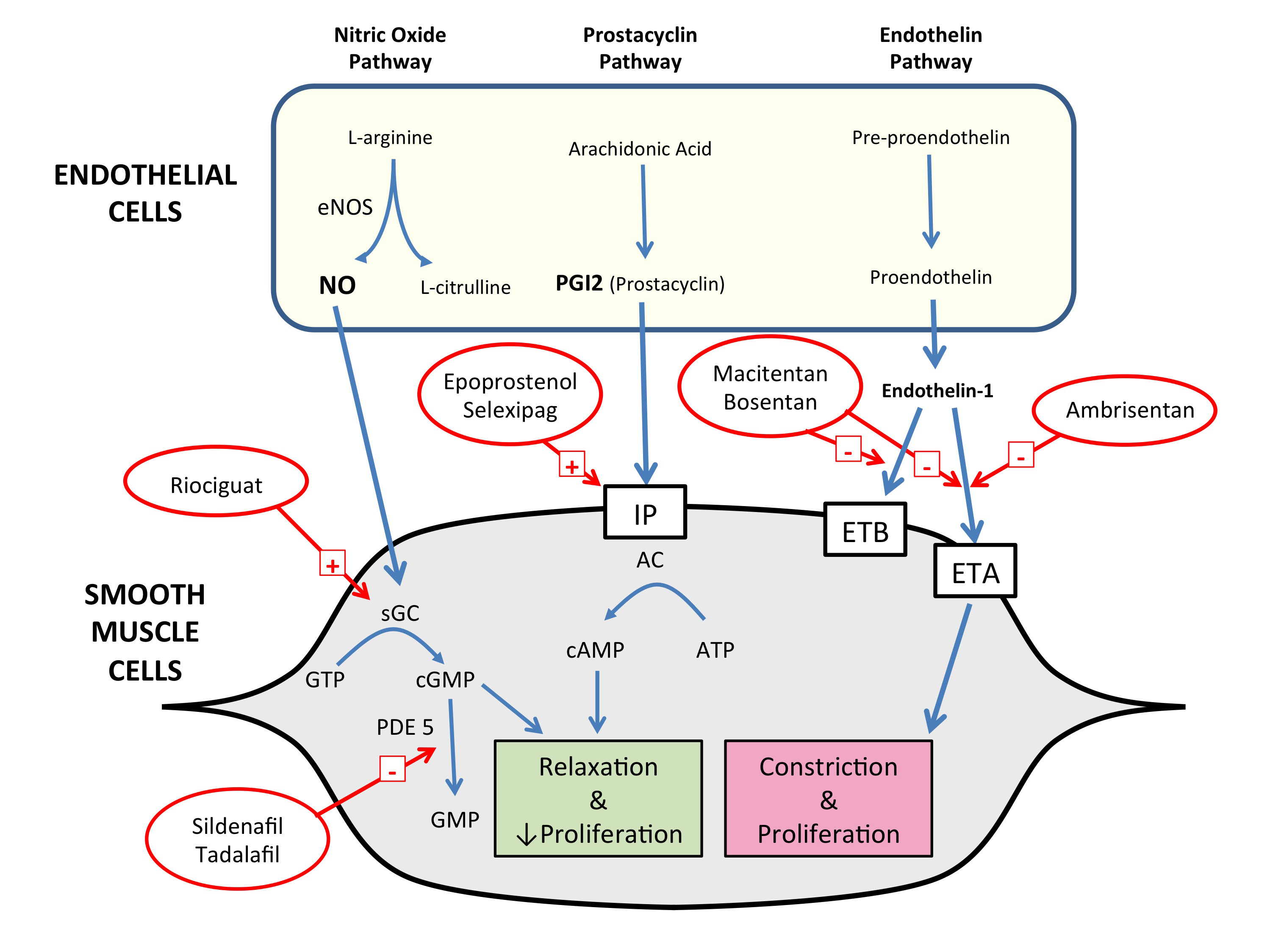
PAH is a disease of endothelial dysfunction and abnormal remodelling of the pulmonary vessels, leading to luminal narrowing, loss of normal vasodilator responses and obliteration of pulmonary arterioles, all resulting in elevated PVR. There is migration and proliferation of vascular smooth muscle cells, appearance of myofibroblasts and increases in extracellular matrix, leading to intimal hyperplasia, medial hypertrophy and adventitial fibrosis. Inflammatory infiltrates are a histological feature of PAH.6 It is thought that in situ thrombosis may also lead to vessel loss.7 It has long been recognised that three major endothelial signalling systems within the vessel walls are dysfunctional in PAH, and these have been the major targets for drug therapies (Box 2). Increased activity of the endothelin system promotes vasoconstriction and vascular smooth muscle cell proliferation.8 A further mechanism important in iPAH is reduction in the synthesis and availability of nitric oxide, which has potent pulmonary vasodilator and antiproliferative effects.9 The endothelial cells in iPAH demonstrate a reduction in prostaglandin I2, another vasodilator with antiproliferative properties.10 While the disease primarily affects blood vessels, it generally results in death due to right heart failure.11

Recently, there has been interest in the factors responsible for initiating and driving the process that results in PAH. The most common cause of familial PAH is mutations in the bone morphogenetic protein receptor type 2 (BMPR2) gene, a member of the transforming growth factor beta (TGF-β) signalling family and thought to be responsible in 80% of cases, although mutations associated with PAH have been described in other genes in the TGF-β superfamily.12 However, there have been many specific BMPR2 mutations identified, with the most common accounting for only 8% of familial PAH, making genetic screening for this disease impractical. Not all carriers of the abnormal genes develop PAH, suggesting additional influences are necessary for phenotypic expression, possibly reflecting a “multiple hit” process, similar to that seen with many cancers.13 It has been shown that BMPR2 is downregulated in other forms of PAH, suggesting that this may also be important in non-inherited forms of PAH. Dysregulated immunity and inflammation may also be important in the development of PAH.14 Patients with PAH have been observed to have elevated levels of serotonin. Disruption of the normal serotonin transporter affects the development of PH in animal models, signifying that the interaction between serotonin and vascular smooth muscle cells may be important in the development of PAH.6,14

Chronic thromboembolic pulmonary hypertension

In a small proportion of patients who experience an acute pulmonary embolism, the thrombus will fail to resolve afterwards. This was initially thought to be true for about 0.5% of patients with pulmonary embolism, but a landmark report suggested almost 4% of patients will develop evidence of CTEPH in the 2 years after a pulmonary embolism.15 Patients with CTEPH may typically present with only partial recovery from a pulmonary embolism, followed by a (“honeymoon”) period of stability (which may last months or even years), then subsequent decline. At least 25% of patients have no history of an acute embolus, presenting with progressive breathlessness.4 Although increased propensity to thrombosis seems to predispose to CTEPH (32% of patients in a large international registry had a thrombophilic disorder),4 the underlying abnormality appears to be related to abnormal thrombin or abnormal thrombinolysis — a clot forms in the pulmonary artery but is incompletely broken down, resulting in a complex obstructing lesion in the artery.

Survival at 3 years without surgery is 70%, but this rises to 89% if surgery is performed.16 Although no randomised controlled trial (RCT) has been conducted, pulmonary endarterectomy (PEA), where feasible, is regarded as the treatment of choice, and initial investigations should focus on suitability for PEA. The presence of PH may be evident on echocardiography, but confirmation requires right heart catheterisation.17 Persistent segmental or larger unmatched perfusion defects seen on a nuclear ventilation–perfusion (VQ) scan are highly suggestive of operable CTEPH (a negative VQ scan virtually excludes operable CTEPH, except where the pulmonary artery trunk or both the left and right pulmonary arteries have high grade obstruction).17 A negative computed tomography pulmonary angiography scan does not exclude operable CTEPH and, as such, is not the best screening modality. Pulmonary angiography is currently the gold standard for assessing the extent and surgical accessibility of the obstructing lesions.18

PEA performed in experienced centres carries a surgical mortality of 2–5%.19,20 Most patients return to near normal functional capacity (New York Heart Association [NYHA] Class I or II) after surgery.21 PEA involves extensive endarterectomy from the main pulmonary artery down to segmental branches. It is performed under hypothermic circulatory arrest (18°C) in a few expert centres in Australia.

In patients with persistent PH after PEA, or in those unsuitable for surgery, selective pulmonary vasodilators may be useful. For most selective vasodilators, case series have shown at least haemodynamic improvements. In a 16-week RCT, riociguat (a novel soluble guanylate cyclase stimulator) resulted in haemodynamic improvements, as well as a significant increase in 6-minute walk distance (6MWD) of 46 m.22 The 6MWD is a simple test performed under standardised conditions of the distance walked over flat ground in 6 minutes; it is often used in PH trials. Concern remains that patients who are suitable for surgery should not be initially offered vasodilator therapy, as both the drug and the delay to PEA may lead to an inferior outcome.17 Pulmonary artery balloon angioplasty may offer a viable alternative to PEA in some circumstances, where several discrete, more distal lesions are inaccessible surgically. However, the role of balloon angioplasty as a substitute for PEA is currently unclear, especially in older patients who are less well suited to, or accepting of, major surgery. More systematic study of this matter is warranted.23

Current drugs for pulmonary arterial hypertension

The existing treatments for iPAH target signal pathways proven to be involved in the disease’s pathophysiology (Box 2). Ultimately, normal life expectancy and normal quality of life are the objectives of therapy. Realistically, moving patients to a low risk of mortality with, at worst, mild functional limitation (NYHA Class I–II) is the present target of therapy.24

Patients whose PH may be Group 1 or Group 4 in aetiology should be referred to a specialised PH centre for diagnosis and treatment. Patient education and use of expert centre support are essential for optimal outcomes and minimal side effects using currently available drug treatments (Box 3).

Calcium channel blockers

There is a small group of patients with iPAH (6%) who appear to respond well to high doses of dihydropyridine calcium channel blockers and who may have durable benefit from this therapy.25 These patients are identified using a vasodilator challenge at the time of right heart catheterisation. Patients with no acute vasodilator response should not be treated with high dose calcium channel blockers.

Endothelin receptor antagonists

The oral endothelin receptor antagonists (ERAs) bosentan and ambrisentan (and the newer ERA macitentan) inhibit the signal pathway by blocking endothelin receptors. Monotherapy with bosentan and ambrisentan has produced improvements in 6MWD, PVR and time to clinical deterioration.26,27

Phosphodiesterase type 5 inhibitors

Phosphodiesterase type 5 (PDE5) breaks down cyclic guanosine monophosphate (cGMP) and therefore decreases the vasodilator effects of nitric oxide. The oral PDE5 inhibitors sildenafil and tadalafil decrease cGMP degradation, which increases the activity of the nitric oxide system and clinically improves 6MWD in patients with iPAH.28,29 Tadalafil also increases time to clinical deterioration compared with placebo.29

Prostanoids

The two major prostanoids used in Australia are epoprostenol and iloprost. Epoprostenol is the most effective single agent for treatment of PAH, but it requires delivery of a continuous intravenous infusion via a central venous catheter. It has been shown to improve 6MWD, haemodynamics, quality of life and survival.30 Recently, a more thermostable formulation of epoprostenol has become available, which may be more convenient to use as it eliminates the need for a cold pack to maintain the drug’s temperature at the required level. Inhaled iloprost improves functional class, 6MWD and time to clinical deterioration,31 but requires up to nine inhalations per day.

Anticoagulants

Early pathological studies showed thrombosis within small lung vessels in patients with PAH,32 leading to treatment with anticoagulants becoming part of standard therapy. Anticoagulation in patients with iPAH may be of benefit, with some observational series indicating improvements in survival, but uncertainty persists due to the low level of evidence.7,25,33 Use of anticoagulation in patients with PAH and scleroderma is more controversial.34 Recent guidelines for the treatment of iPAH state that oral anticoagulation with warfarin may be considered in treatment algorithms.35

New drugs for pulmonary arterial hypertension

While calcium channel blockers, intravenous epoprostenol, the oral ERAs bosentan and ambrisentan, and PDE5 inhibitors sildenafil and tadalafil are well established in the treatment of iPAH in Australia, new drugs have recently been introduced or will soon be available (Box 3). Direct comparison with currently available treatments is difficult, as most current treatments were tested in relatively small trials of short duration, using surrogate endpoints based largely on haemodynamic or functional measures. In contrast, trials of newer agents have been larger and in many cases have used mortality and morbidity endpoints, such as time to clinical deterioration, to demonstrate efficacy. Direct comparison is also difficult due to the widespread use of background PAH therapies in recent studies.

Macitentan

Macitentan is a recently introduced non-selective ERA developed with the aims of improving effectiveness and reducing the side effects, toxicities and drug interactions associated with earlier ERAs. Macitentan has been assessed in the SERAPHIN trial, the largest RCT of ERAs, in which two macitentan doses (3 mg and 10 mg) were compared with placebo in 742 patients with Group 1 PH over a median treatment period of 115 weeks.36 Occurrence of the primary outcome (first occurrence of a composite of death, lung transplantation, atrial septostomy, initiation of intravenous or subcutaneous prostanoids, or worsening of PAH) was reduced by 30% (P = 0.01) with 3 mg and by 45% (P < 0.001) with 10 mg of macitentan. This was mostly driven by a reduction in occurrence of worsening of PAH. Worsening of PAH was defined as a decrease in 6MWD of at least 15%, worsening of PAH symptoms and the need for additional PAH treatment. Reductions in hospitalisation were also seen with macitentan treatment. About two-thirds of patients in this study were already taking other PAH treatments, predominantly PDE5 inhibitors, with the remaining third taking the study drug as monotherapy. Improved outcomes with macitentan were seen in both the treatment-naive patients and those using combination therapy.36

Riociguat

The effectiveness of PDE5 inhibitors may be limited because PAH is a state of nitric oxide deficiency, and inhibiting breakdown of the already reduced levels of cGMP may not be the most effective strategy. Riociguat acts directly to stimulate soluble guanylate cyclase and thus increase cGMP independent of nitric oxide levels.37 Riociguat has been tested in patients with Group 1 PH in the PATENT-1 trial.38 This study compared two doses of riociguat with placebo in 443 patients, of whom half were treatment naive. Most of those taking the active drug were treated with 2.5 mg three times a day (254 patients). Riociguat resulted in a placebo-corrected increase in 6MWD of 36 m after 12 weeks of treatment (P < 0.001). In addition, improvements were seen in PVR, time to clinical deterioration, functional class, N-terminal pro-brain natriuretic peptide levels and quality of life with riociguat treatment.38

Selexipag

Although intravenous epoprostenol is a highly effective treatment for PAH, the logistics and infective complications associated with chronic intravenous administration limit its use.30 Other intravenous, oral and subcutaneous prostanoids have been limited by difficulties in their administration or intolerable side effects.31,39 More recently, a non-prostanoid prostacyclin receptor agonist, selexipag, has been studied in patients with PAH. The 1152 patients with Group 1 PH in the event-driven GRIPHON trial were randomly allocated to receive selexipag or placebo and followed for a median of 70.7 and 63.7 weeks, respectively.40 About 80% were already using specific PAH therapies, and the remaining 20% were treatment naive. Selexipag was uptitrated to 1600 μg twice daily or the highest tolerated dose. Selexipag treatment was associated with a 40% lower risk of the primary composite endpoint of death, hospitalisation for PAH, disease progression, initiation of parenteral prostanoid or long term oxygen therapy, or lung transplantation. The magnitude of benefit did not differ across different achieved doses, suggesting that the maximum tolerated dose is the correct dose for the patient. Several side effects, including jaw pain, nausea, diarrhoea, vomiting and myalgia, were more common in those treated with selexipag.

Combination therapy

In diseases such as cancer, human immunodeficiency virus, systemic hypertension and heart failure, treatment with combinations of drugs has proven to be more effective than treatment with a single agent. There is now evidence that combining specific therapies for PAH is more effective than monotherapy. Combination therapy may be used either sequentially or as initial therapy at the time of diagnosis. The strongest evidence for initial combination therapy comes from the AMBITION trial, which showed that an initial combination of ambrisentan and tadalafil was superior to either agent alone, with a 50% reduction in the occurrence of a primary endpoint event (first event of clinical failure) for those taking combination therapy compared with the monotherapy group.41 There were also beneficial effects on secondary endpoints.

In recent trials of new therapies, the rate of background PAH therapy has been significant, with about 80% of patients in the GRIPHON trial of selexipag,40 50% of patients in the PATENT-1 trial of riociguat,42 and 64% of patients in the SERAPHIN trial of macitentan36 having the trial drug added to background PAH therapy. Benefit has been observed in both those taking background therapy and treatment-naive patients, suggesting new drugs should be added to existing treatments, rather than substituted for them; a position that is supported by current international guidelines and consensus statements.35,43 A small pilot study in France showed excellent outcomes with initial triple combination therapy, including an intravenous prostenoid.44 A large study of initial combination double versus triple therapy (TRITON), comparing macitentan and sildenafil with selexipag or placebo, will commence recruitment in the next few months and we hope will help define the role for more aggressive initial oral therapies.

Access to treatment in Australia

There have been no head-to-head clinical trials of sufficient size or quality to make any scientifically based judgement on which of the available drugs can be considered first-line therapy for PAH, and local practice around the world is often based on cost. It is also not known how many patients in Australia are using each type of therapy, as the databases that exist likely do not reflect the whole country.

Two issues affect patient access to specific PAH therapies in Australia. While recent trial data indicate that combination therapy is superior to monotherapy, currently only treatment with one agent at a time is funded through the Pharmaceutical Benefits Scheme (PBS). However, based on our experience, we are aware that a significant number of Australian patients are using more than one PAH treatment, some of which is self-funded, some funded through hospitals and some sourced through compassionate access schemes. There are no systematic data on the use of combination therapy by patients.

Recent changes to the PBS have simplified the ongoing treatment of patients with PAH, with a reduction in administrative and repeat testing burden. Patients who have commenced PAH treatment or who have changed drugs must demonstrate disease stability (as described in the PBS explanatory notes) or improvement using 6MWD and echocardiography and/or right heart catheterisation after 6 months of treatment. Ongoing treatment at 12 months and beyond will no longer require demonstration of stability. Repeat assessment should, however, continue to be performed on clinical grounds and in accordance with practice guidelines.35

Future treatments

Present therapies rarely induce remission and never cure, to use a cancer analogy, and overall 3-year survival with their use is about 75%.36 Alternative treatment pathways identified through basic research are being evaluated. Initial enthusiasm for the tyrosine kinase inhibitor imatinib has been dampened by the high rate of side effects in a clinical trial setting.45 Several other compounds are in pre-clinical or early clinical evaluation.46

For patients whose PAH is not well controlled using available therapies, transplantation (double lung or heart–lung) remains an option. With improvements in organ availability in Australia, this is both feasible and likely; however, chronic lung allograft dysfunction still limits median survival to about 8 years.47 Developments in regenerative medicine may allow repopulation of a donated lung (rendered acellular) with autologous stem cells,48 an exciting approach currently in pre-clinical development.

The recognition that up to 30% of patients with apparently idiopathic PAH have an identified gene defect raises the prospect of gene therapy.49 Mutations of the BMPR2 gene are the commonest, and correcting this defect with gene therapy has been suggested — using an adenovirus, a vector can transfect the relevant cell.50 However, this leads to an immunological response, which means this approach will be unsuitable for recurrent treatments. Endothelial progenitor cells may help repair the vascular endothelium, and there are data from human studies on this approach.51 More recently, the use of endothelial progenitor cells as vectors for gene therapy has been proposed, with a human study recently attempting to introduce an inducible nitric oxide synthase (iNOS) gene into the pulmonary vascular endothelium.52

Conclusion

The past 10 years have seen the introduction into routine clinical practice of new oral and intravenous therapies for PAH, based on the endothelin, nitric oxide and prostacyclin pathways, leading to improvements in functional capacity and survival. Emerging evidence supports more widespread use of combination therapy, as both initial and sequential treatment, although there are significant barriers to this use in Australia. CTEPH remains an important cause of PH to identify because of its potential for successful treatment using surgery, medication and possibly angioplasty. There is enthusiasm for exploring new pathways that are important in PAH to identify new agents that may address more basic elements of pathophysiology and arrest or reverse the process of pulmonary blood vessel obstruction and obliteration.

Box 1 –
Simplified World Health Organization classification of pulmonary hypertension*

  1. 1. Pulmonary arterial hypertension (PAH)
    1. 1.1 Idiopathic PAH
    2. 1.2 Heritable PAH (BMPR2, ALK1, ENG, CAV1 and other mutations)
    3. 1.3 Drug- and toxin-induced pulmonary hypertension
    4. 1.4 PAH associated with:
      1. 1.4.1 Connective tissue disease
      2. 1.4.2 HIV infection
      3. 1.4.3 Portal hypertension
      4. 1.4.4 Congenital heart disease

  2. 1′. Pulmonary veno-occlusive disease (PVOD) and/or pulmonary capillary haemangiomatosis (PCH)
  3. 2. Pulmonary hypertension due to left heart disease
    1. 2.1 Left ventricular systolic dysfunction
    2. 2.2 Left ventricular diastolic dysfunction
    3. 2.3 Valvular disease
    4. 2.4 Congenital or acquired left ventricular outflow tract obstruction and congenital cardiomyopathies
  4. 3. Pulmonary hypertension due to lung disease and/or hypoxia
    1. 3.1 Chronic obstructive pulmonary disease
    2. 3.2 Interstitial lung disease
    3. 3.3 Mixed obstruction and restriction
    4. 3.4 Sleep disordered breathing
    5. 3.5 Chronic exposure to high altitude
  5. 4. Chronic thromboembolic pulmonary hypertension
  6. 5. Pulmonary hypertension due to unclear multifactorial mechanisms

* Adapted from Simonneau et al.2 † The classification group 1’ was created because PVOD and/or PCH share many characteristics with idiopathic PAH but are recognised to also have distinct differences.

Box 2 –
Diagrammatic representation of the key abnormal pathways targeted in treatment of pulmonary arterial hypertension and the mode of action of current and new drugs


AC = adenylate cyclase. ATP = adenosine triphosphate. cAMP = cyclic adenosine monophosphate. cGMP = cyclic guanosine monophosphate. eNOS = nitric oxide synthase. ET = endothelin. GTP = guanosine triphosphate. IP = IP receptor. NO = nitric oxide. PDE5 = phosphodiesterase type 5. sGC = soluble guanylate cyclase.

Box 3 –
Drug treatments for idiopathic pulmonary arterial hypertension (PAH)

Class of drug


Endothelin receptor antagonists

PDE5 inhibitors

Prostanoids

Calcium channel blockers

sGC stimulators

Prostacyclin receptor agonists


Generic names

Bosentan, ambrisentan, macitentan

Sildenafil, tadalafil

Epoprostenol, iloprost

Nifedipine, amlodipine, diltiazem

Riociguat

Selexipag

Route of administration

Oral

Oral

Intravenous, inhaled, oral

Oral

Oral

Oral

Clinical improvements

6MWD, PVR, TCD, fewer morbidity and mortality events*

6MWD, TCD

6MWD, TCD, haemodynamics, survival, quality of life

Survival, haemodynamics, functional capacity

6MWD, TCD, haemodynamics, functional capacity

Reduced death, hospitalisation and worsening of PAH; haemodynamics

Common side effects

Hepatotoxicity,§ peripheral oedema, nasal congestion, nasopharyngitis,* headache,* anaemia

Headache, dyspepsia, diarrhoea, myalgia, flushing, epistaxis

Central venous access infection or blockage, cough, headache, flushing, jaw pain

Hypotension, peripheral oedema

Headache, dyspepsia, peripheral oedema, dizziness, hypotension

Headache, nasopharyngitis, anaemia, jaw pain, nausea


PDE5 = phosphodiesterase type 5. sGC = soluble guanylate cyclase. 6MWD = 6-minute walk distance. PVR = pulmonary vascular resistance. TCD = time to clinical deterioration. * Macitentan only. † Tadalafil only. ‡ Epoprostenol only. § Bosentan only. ¶ Ambrisentan only.

New developments in coronary stent technology

Current performance standards are high and promising new technologies are finding it difficult to compete

The narrative review in this issue of the MJA by Chen and Jepson1 outlines the enormous advances made in stent technology since Sigwart and colleagues published the first report on the clinical use of coronary stents in 1987.2 Driven by technological progress and rigorous scientific study, coronary stents have advanced rapidly to the stage where a patient’s coronary stenosis can be safely and reliably opened.

A 60-year-old man is brought to the emergency department with severe chest pain, his electrocardiogram showing marked anterior ST elevation. He has a ventricular fibrillation arrest and is shocked into sinus rhythm. Catheterisation laboratory staff are on their way and, within minutes of arrival, a coronary angiogram shows complete occlusion of the left anterior descending artery. A guide-wire is inserted and flow is established after balloon inflation. His pain is settling as a drug-eluting stent is inserted. He is discharged on Day 3.

Despite benefiting dramatically from stent technology, our patient’s future is not without risks and challenges. He needs to take dual antiplatelet drugs for 1 year and postpone elective surgery. Over the next 5 years, he faces a stent thrombosis rate of 1.4–4.0%, and an almost 20% chance of adverse events including death, myocardial infarction (MI) or repeat procedures.35 As Chen and Jepson point out in their review, there appears to be an ongoing risk of ischaemic events for some years after successful stent implantation. The cause of this is likely a combination of stent factors including chronic inflammation, uncovered struts and neoatherosclerosis within the stent, as well as progression of atherosclerotic disease elsewhere. In a long term follow-up of patients treated with second generation stents, around 50% of adverse events were not related to the stent but were due to plaque progression at other coronary sites.6 This emphasises the importance of secondary prevention strategies for the long term health and wellbeing of patients treated with stents.

Progress in stent technology has made percutaneous coronary intervention (PCI) a relatively easy and safe procedure with visually impressive results for doctors and patients. Greater adoption of radial artery access has also contributed to the added safety of these procedures.7 It can be difficult to resist the temptation to insert stents in order to obtain aesthetically satisfying results in imperfect coronary arteries. Yet there is evidence from a randomised trial to suggest that PCI in patients with stable coronary disease does not reduce the risk of death, MI or other major cardiovascular events compared with optimal medical treatment.8 The challenge for physicians is not how to place a stent, but deciding when to intervene to produce the best outcomes for patients in a cost-effective way. Assessment of patients for evidence of ischaemia and use of fractional flow reserve measurement in coronary arteries should have a larger influence in decision making than reliance on visual assessment of coronary arteries at the time of angiography.

We also need to better understand the disease that we are treating. It is simplistic to think that stent insertion will remain the optimal treatment for all forms of coronary disease such as thin-cap plaque rupture, fibrous plaque, calcified plaque, thrombus, erosions, spontaneous dissection, long lesions, chronic total occlusions, bifurcation lesions and restenosis. Greater use of intracoronary imaging before and after stent insertion can help us to better diagnose the nature of coronary stenosis, with the future prospect of optimising treatment for individual patients.

Current generation drug-eluting stents have set such a high standard in safety and efficacy for such a wide range of lesions that new technologies will find it difficult to show superiority without large and long term clinical trials. Bioresorbable vascular scaffolds are an example of a very promising technology that is finding it difficult to compete with second generation stents in a wide range of lesion types. Bifurcation stents are another promising advance, but a recent randomised trial showed that a dedicated bifurcation stent was associated with a higher risk of MI compared with a conservative one-stent strategy.9

The success of stent technology has made PCI one of the most commonly performed procedures in medicine today and we look forward to further developments in this field. Yet, ironically, we would also welcome future reports of a decline in stent insertion in Australia, as this would indicate that as a society, we are starting to win the battle against coronary artery disease. With the rising tide of obesity and diabetes, this appears unlikely in the foreseeable future.

[Comment] Stroke and mortality after atrial fibrillation—a global struggle

Atrial fibrillation is a widely recognised health-care challenge with increasing prevalence across the world. Epidemiological observations mainly attribute this increase to an ageing population and better prognosis in distinguishing it from other cardiac disorders such as myocardial infarction.1 Research into atrial fibrillation has focused on antithrombotic management for stroke prevention and mortality, in the era of non-vitamin K antagonist oral anticoagulants.2 However, although these large, phase 3 trials enrolled patients from many centres in different countries and included broad populations, regional differences and between-centre variations in quality of treatment might have affected the results.

Development of the Canadian Syncope Risk Score to predict serious adverse events after emergency department assessment of syncope [Research]

Background:

Syncope can be caused by serious conditions not evident during initial evaluation, which can lead to serious adverse events, including death, after disposition from the emergency department. We sought to develop a clinical decision tool to identify adult patients with syncope who are at risk of a serious adverse event within 30 days after disposition from the emergency department.

Methods:

We prospectively enrolled adults (age ≥ 16 yr) with syncope who presented within 24 hours after the event to 1 of 6 large emergency departments from Sept. 29, 2010, to Feb. 27, 2014. We collected standardized variables at index presentation from clinical evaluation and investigations. Adjudicated serious adverse events included death, myocardial infarction, arrhythmia, structural heart disease, pulmonary embolism, serious hemorrhage and procedural interventions within 30 days.

Results:

We enrolled 4030 patients with syncope; the mean age was 53.6 years, 55.5% were women, and 9.5% were admitted to hospital. Serious adverse events occurred in 147 (3.6%) of the patients within 30 days after disposition from the emergency department. Of 43 candidate predictors examined, we included 9 in the final model: predisposition to vasovagal syncope, heart disease, any systolic pressure reading in the emergency department < 90 or > 180 mm Hg, troponin level above 99th percentile for the normal population, abnormal QRS axis (< –30° or > 100°), QRS duration longer than 130 ms, QTc interval longer than 480 ms, emergency department diagnosis of cardiac syncope and emergency department diagnosis of vasovagal syncope (C statistic 0.88, 95% confidence interval [CI] 0.85–0.90; optimism 0.015; goodness-of-fit p = 0.11). The risk of a serious adverse event within 30 days ranged from 0.4% for a score of –3 to 83.6% for a score of 11. The sensitivity was 99.2% (95% CI 95.9%–100%) for a threshold score of –2 or higher and 97.7% (95% CI 93.5%–99.5%) for a threshold score of –1 or higher.

Interpretation:

The Canadian Syncope Risk Score showed good discrimination and calibration for 30-day risk of serious adverse events after disposition from the emergency department. Once validated, the tool will be able to accurately stratify the risk of serious adverse events among patients presenting with syncope, including those at low risk who can be discharged home quickly.

Disparities in acute in-hospital cardiovascular care for Aboriginal and non-Aboriginal South Australians

The known Disparities in the treatment of Aboriginal and non-Aboriginal patients hospitalised with acute coronary syndromes have been reported. 

The new After adjusting for age and other factors, Aboriginal status was independently associated with lower coronary angiography rates. Angiography was more likely if family members or Aboriginal liaison officers were present. Revascularisation rates and prescription of medical therapies were similar for Aboriginal and non-Aboriginal patients who had undergone angiography. 

The implications The reasons for lower angiography rates among Aboriginal patients are complex, but equality of treatment can be achieved. Improving the hospital experience for Aboriginal patients is needed to reduce disparities in treatment. 

Coronary heart disease (CHD) contributes significantly to the 10-year life expectancy gap between Aboriginal and non-Aboriginal Australians.13 CHD mortality is estimated to be twice as high among Aboriginal Australians,3 accounting for 14% of all deaths of Aboriginal people.4 A higher incidence of acute coronary syndromes (ACS), particularly of acute myocardial infarctions (AMI), among Aboriginal Australians is a major contributor to premature mortality in this population. In 2006, the Australian Institute of Health and Welfare identified major disparities in the management of Aboriginal and Torres Strait Islander patients hospitalised with ACS between 2002 and 2003, including a 40% lower rate of coronary angiography, a 40% lower rate of percutaneous coronary intervention (PCI), and a 20% lower rate of coronary artery bypass graft surgery (CABG).5

Given the extent of the inequalities related to cardiovascular disease, there is clearly a need to identify and overcome problems that contribute to these health disparities. To expand knowledge in this area, our study examined the in-hospital management of Aboriginal people with an ACS, focusing on explaining these disparities. Our aims were: (i) to assess differences in the rates of angiography and subsequent revascularisation for Aboriginal and non-Aboriginal South Australians presenting with an ACS, taking into account age and other factors; and (ii) to explore the reasons for any disparities by undertaking a detailed review of individual hospital admissions.

Methods

This was an observational study of Aboriginal and non-Aboriginal patients presenting with an ACS to any public tertiary hospital in South Australia between January 2007 and December 2012. We undertook a retrospective analysis of hospital administrative data and a chart review of all admissions of Aboriginal people with an ACS during this period (matched with a control cohort of non-Aboriginal patients).

Establishment of the expert working group

Following the release of Better hospital care for Aboriginal and Torres Strait Islander people experiencing heart attack,6 it was recognised that contemporary SA-specific data were required for evaluating disparities in care in this state. Consequently, the Heart Foundation in SA, in collaboration with the SA Health Cardiology Clinical Network, established a research team to provide the data. The project was overseen by an expert working group with representation from the research team and each SA Health local health service, as well as Aboriginal health professionals.

Administrative data

To investigate differences in diagnostic angiography and revascularisation (PCI/CABG) rates in Aboriginal and non-Aboriginal people, hospital admissions for ACS to all South Australian public tertiary facilities, with separations between 1 January 2007 and 31 December 2012, were obtained from the Integrated South Australian Activity Collection (ISAAC). ISAAC records are coded using International Statistical Classification of Diseases and Related Health Problems, 10th revision, Australian modification (ICD-10-AM) codes. All records were based on separations (episodes of care) rather than individual patients. Records were included in the analysis if the principal diagnosis was AMI (ICD-10-AM, I21.x) or unstable angina (I20.x) and the admission was recorded as an acute episode of care with casualty/emergency department as the source of referral. To avoid contamination of the dataset, records for interhospital transfers were excluded. Records in which Indigenous status was not stated or was inadequately described were also excluded.

Chart review

To further examine the in-hospital management of Aboriginal patients with an ACS, a chart review was undertaken, using 1:1 matching and a standardised case report form. The hospital medical records for each separation recorded in the administrative data of patients who were identified as Aboriginal or Torres Strait Islander were extracted. They were matched as closely as possible with the next admission to the same hospital of a non-Aboriginal patient of the same age (within 10 years) and sex, and with the same principal diagnosis. Documentation of the medical decision-making process about whether the patient received invasive management was also reviewed; influences on this process were categorised as clinical or non-clinical (ie, patient-related factors).7 Data quality for the chart review was ensured by using trained abstractors and record re-abstraction for 10% of cases. Training included a detailed data dictionary and coding instructions, a testing phase with training examples, and individual education sessions on site before and during data collection.

Statistical analysis

Administrative data

Age at admission (mean, standard deviation) for Aboriginal and non-Aboriginal patients was compared with the Student t test. Comparisons of categorical endpoints for Aboriginal and non-Aboriginal patients are presented as percentages, and were assessed by logistic regression. Unadjusted and age-adjusted comparisons are reported as odds ratios with 95% confidence intervals (CIs). The primary outcomes included receiving diagnostic coronary angiography and, for patients for whom angiography was performed, PCI and CABG rates. To explore the association between invasive management and Aboriginal status, logistic regression models were fitted for each outcome. Modelling commenced with fully saturated models that included Aboriginal status and all remaining predictors with P < 0.20 as factors; non-significant variables (P ≥ 0.05) were then systematically removed from the models.

Chart review

Clinical outcomes for Aboriginal and non-Aboriginal patients are presented as percentages and compared by logistic regression as described above. Symptom duration in minutes (median, interquartile range [IQR]) was compared with the Wilcoxon rank-sum test. The primary outcome in the chart review was the prevalence of clinical and non-clinical factors associated with non-invasive management; outcomes for the two groups were compared by logistic regression, with unadjusted and age-adjusted results reported.

All tests were two-tailed (α = 0.05). All analyses were performed in Stata/IC 11.2 for Mac.

Ethics approval

This study was approved by the SA Health Human Research Ethics Committee (reference, HREC-13-SAH-89; 461/07/2014) and the Aboriginal Health Research Ethics Committee (reference, 04-13-516).

Results

Administrative data

During January 2007 – December 2012, a total of 13 843 separations with a principal diagnosis of ACS were recorded. Aboriginal status was not stated or inadequately described in 722 records (5.6%), leaving 13 071 admissions records for analysis, of which 274 (2.1%) referred to Aboriginal patients. The mean age of the Aboriginal patients was about 16 years lower than for non-Aboriginal patients, and a higher proportion were women (Box 1). The age distribution by Aboriginal status and sex is summarised in Appendix 1. Most patients presented with an AMI; the figure was similar for the two groups (57%), despite the younger age profile of the Aboriginal patients. Evaluation of additional diagnosis codes found a higher prevalence among Aboriginal patients of several cardiovascular risk factors and comorbidities, including diabetes, smoking, dyslipidaemia and renal failure (Box 1).

Analysis of hospital procedure codes indicated that 6069 separations (46.4%) included a diagnostic coronary angiogram during the admission for an ACS (Box 2). The proportion was similar for Aboriginal and non-Aboriginal patients, but age-adjusted analyses identified that Aboriginal patients were significantly less likely to undergo the procedure (Box 2). Box 3 shows the prevalence of coronary angiography by age group, indicating a significant disparity for Aboriginal patients in the 45–54 years (P < 0.001) and 55–64 years (P = 0.001) age groups.

For the entire cohort, patients who underwent coronary angiography were significantly younger than those who did not (mean [SD], 64.0 years [12.9] v 73.8 years [13.8]; P < 0.001), and a greater proportion were men (72% v 58%; odds ratio [OR], 1.87; 95% CI, 1.74–2.01; P < 0.001). Multivariable logistic regression identified eight independent factors significantly associated with angiography: Aboriginal status (OR, 0.4; 95% CI, 0.3–0.5; P < 0.001); age (as a continuous variable: OR [for each one-year increase in age], 0.9; 95% CI, 0.9–1.0; P < 0.001); AMI as principal diagnosis (OR, 5.8; 95% CI, 5.3–6.3; P < 0.001); sex (OR [men v women], 1.3; 95% CI, 1.2–1.4; P < 0.001); major cities (OR [v outer regional/remote/very remote area residence], 2.9; 95% CI, 2.3–3.7; P < 0.001); renal failure (OR, 0.5; 95% CI, 0.4–0.6; P < 0.001); heart failure (OR, 0.5; 95% CI, 0.5–0.6); P < 0.01); and airway disease (OR, 0.6; 95% CI, 0.5–0.7; P < 0.001).

After adjustment for age, the odds of Aboriginal patients undergoing PCI were significantly lower than for non-Aboriginal patients (Box 2). The between-groups odds ratios for undergoing CABG were significant in neither the unadjusted nor the age-adjusted analyses. However, when the analysis of interventional procedures was restricted to patients who underwent coronary angiography, PCI rates for Aboriginal and non-Aboriginal patients were similar, as were those of CABG and revascularisation overall (Box 2). For admissions that included a coronary angiogram, multivariable models assessing independent predictors of revascularisation (PCI, CABG) did not find an association with Aboriginal status.

Chart review

We reviewed medical record abstracts for 274 Aboriginal and 274 non-Aboriginal patients. Aboriginal patients were matched for sex with non-Aboriginal patients (57% were men), but were significantly younger (mean age [SD], 53.1 years [10.5] v 59.0 [10.4] years; P < 0.001). The number who arrived at hospital by ambulance was similar for Aboriginal and non-Aboriginal patients (148 [61%] v 149 [60%]; age-adjusted OR, 1.3; 95% CI, 0.9–1.8; P = 0.24), as was symptom duration (median time before hospital presentation [IQR], 160 min [80–415 min] v 165 min [80–419 min]; P = 0.62). The angiography rate among Aboriginal patients was lower than for non-Aboriginal patients in both the unadjusted and age-adjusted analyses (141 [51%] v 169 [62%]; age-adjusted OR, 0.5; 95% CI, 0.4–0.7; P < 0.001). Stratification according to the National Heart Foundation/Cardiac Society of Australia and New Zealand ACS guidelines8 indicated a similar risk burden for the patient groups (Appendix 2). The proportion of Aboriginal patients who underwent angiography was lower in each risk group, but the difference was statistically significant only for patients with high risk non-ST-elevation acute coronary syndrome (NSTEACS; 49% [59 patients] v 60% [64 patients]; age-adjusted OR, 0.5; 95% CI, 0.3–0.9; P = 0.02) (Box 4). Aboriginal patients who received care facilitated by an Aboriginal liaison officer were significantly more likely to have an angiogram (28% [27 patients] v 9% [10 patients]; OR, 3.9; 95% CI, 1.8–8.6; P = 0.001), as were Aboriginal patients who arrived at the hospital with an escort (43% [32 patients] v 24% [25 patients]; OR, 2.4; 95% CI, 1.3–4.6; P = 0.01).

The documentation on the decision not to proceed with angiography for Aboriginal and non-Aboriginal patients is summarised in Box 5. Non-clinical factors were more frequently cited to explain managing an ACS without angiography in documentation for Aboriginal patients than for non-Aboriginal patients. For more than one third of Aboriginal patients, the reason for choosing conservative management was unclear, compared with 10% for non-Aboriginal patients (Box 5).

At discharge, the prescription of guideline-recommended therapies for AMI patients was similar for Aboriginal and non-Aboriginal patients (Box 6). Regardless of Aboriginal status, secondary prevention therapies, including aspirin, β-blockers and cardiac rehabilitation referral, were prescribed significantly less frequently for those who did not undergo coronary angiography than for patients who did (Appendix 3).

Discussion

In this evaluation of patients attending the major tertiary facilities in SA for the treatment of ACS, the mean age of Aboriginal patients was about 15 years lower than for non-Aboriginal patients. Despite their being younger, high risk features were worryingly common, including higher rates of background cardiovascular risk factors and comorbidities. Importantly, we identified that the major difference in the in-hospital treatment of Aboriginal patients with an ACS was the rate of coronary angiography: after correcting for the effects of age, sex, principal diagnosis, comorbidities and remoteness, Aboriginal patients with an ACS were significantly less likely to undergo this diagnostic procedure. However, we also found that revascularisation (PCI or CABG) rates following angiography were similar for Aboriginal and non-Aboriginal patients. Similar to findings in the Northern Territory,9 Aboriginal and non-Aboriginal patients who had undergone angiography were prescribed evidence-based therapies on discharge at comparable rates. These findings show that equivalent treatment for Aboriginal and non-Aboriginal patients is achievable.

The lower age-adjusted odds of Aboriginal patients undergoing diagnostic angiography is concerning, but consistent with previous reports of lower rates of cardiac intervention in national5 and state-based datasets.10 Similar rates of CABG, but not of PCI, among Aboriginal and non-Aboriginal patients have been described.10 We, however, found similar rates of PCI for the two population groups; the reasons for this difference are unclear, but may include advances in PCI technology11 that have expanded the range of Aboriginal patients who can be treated with this intervention. Unlike other studies, we also undertook a restricted analysis of revascularisation rates that included only admissions where a diagnostic angiogram had been performed, thereby differentiating different sources of disparity; this analysis indicated that the major difference involved the decision about diagnostic angiography. Research in the United States exploring racial disparities in treating CHD also found that the greatest disparity was the referral to angiography.12,13

To further examine the causes of the disparity in angiography rates, we undertook a medical record review of all Aboriginal ACS admissions in the hospital data, matched with data for a non-Aboriginal cohort. This information provided insight into the medical decision-making processes for Aboriginal ACS patients and confirmed that a negative hospital experience, bias based on complex comorbidities or presumed adherence to medications that favoured conservative management, and patient choice were implicated in the difference in angiography rates.6,14,15

In 56% of cases in which Aboriginal patients did not undergo angiography, the decision was attributed to patient-related factors or no clear justification was provided, compared with 17% for non-Aboriginal patients. The rate of discharge against medical advice was high among Aboriginal patients who did not receive angiography (10.5%). This raises concerns about barriers to quality care for Aboriginal people, including poor engagement and communication, a lack of coordinated care, and inadequate cultural competence of health care providers.16,17 All of these factors can result in isolation, fear and disengagement by the patient.9

The importance of shared discussions and cultural support structures is highlighted by our findings, as Aboriginal patients who arrived at the hospital with an escort (family member or friend) were more likely to undergo angiography. Encouraging shared decision making and enabling systems that support a companion during the hospital stay are crucial for improving Aboriginal health care.14 Our data also strongly support the involvement of Aboriginal liaison officers, as their presence was associated with an increased likelihood of angiography. Although we cannot presume a direct causative relationship, these interactions are understood to improve communication and coordination, and to assist in alleviating fear.17

It has previously been reported that Aboriginal people with symptoms suggestive of a heart attack may delay presenting to a hospital.9 However, our findings suggest this situation may be improving, as symptom duration at hospital presentation was similar for Aboriginal and non-Aboriginal patients. Strategies that target early response and action may now be having an impact, with a number of culturally appropriate health resources and education tools available to patients and health care providers.

The major limitations to our study were that the data were drawn from one Australian state, and that the assessed records were based on hospital separations rather than individual patients. Further, patients receiving care in non-tertiary facilities were not included in our analysis. An evaluation of these patients, with a particular focus on comparing patients who were subsequently transferred with those who remained in a non-catheterisation facility, would be desirable. Our investigation could thus be strengthened by broader population coverage and by using linked data.

Our study provides important insights into the in-hospital treatment of Aboriginal patients with an ACS. We found a significant health disparity in the rates of coronary angiography for Aboriginal and non-Aboriginal patients that is not explained by the frequency of complex comorbidities in these populations. Hospitalisation for acute cardiovascular care can be distressing, and for Aboriginal patients this problem is compounded by limited understanding by health care workers of factors influencing Aboriginal health, leading to miscommunication that may reinforce negative perceptions, patient disengagement, and fear. In our study, engagement with the patient’s family and care facilitated by Aboriginal liaison officers each had positive impacts. While there are other contributing factors to disparities in treatment, health care workers and systems that facilitate a constructive hospital experience will improve the ability to provide effective care for Aboriginal patients.

Box 1 –
Clinical characteristics of Aboriginal and non-Aboriginal patients presenting with an acute coronary syndrome to South Australian tertiary hospitals, 2007–2012

Aboriginal patients

Non-Aboriginal patients

Odds ratio for Aboriginal patients


Unadjusted (95% CI)

P

Age-adjusted (95% CI)

P


Total number of separations

274

12 797

Age at admission (years), mean (SD)

53.1 (10.5)

69.6 (14.1)

< 0.001

Sex (women)

117 (43%)

4460 (35%)

1.4 (1.1–1.8)

0.01

2.7 (2.1–3.5)

< 0.001

Location*

Major cities

203 (74%)

10 730 (84%)

0.6 (0.4–0.7)

< 0.001

0.8 (0.6–1.1)

0.19

Inner regional

12 (4.4%)

1615 (13%)

0.3 (0.2–0.6)

< 0.001

0.2 (0.1–0.4)

< 0.001

Outer regional

25 (9.2%)

310 (2.4%)

4.1 (2.6–6.2)

< 0.001

2.6 (1.7–4.0)

< 0.001

Remote/very remote

33 (12.1%)

128 (1.0%)

14 (9.1–20)

< 0.001

9.2 (5.9–14)

< 0.001

Principal diagnosis

Unstable angina

115 (42%)

5403 (42%)

0.9 (0.7–1.2)

0.93

0.9 (0.8–1.3)

0.51

Acute myocardial infarction (NSTEMI or STEMI)

155 (57%)

7241 (57%)

1.0 (0.8–1.3)

0.99

1.0 (0.8–1.3)

0.71

Unspecified acute myocardial infarction

4 (1%)

153 (1%)

1.2 (0.5–3.3)

0.69

2.3 (0.8–6.5)

0.11

Cardiovascular risk factors

Hypertension

162 (59%)

7745 (61%)

0.9 (0.7–1.2)

0.64

1.3 (1.0–1.7)

0.20

Diabetes

121 (44%)

2737 (21%)

2.9 (2.3–3.7)

< 0.001

3.0 (2.4–3.9)

< 0.001

Current smoker

129 (47%)

2610 (20%)

3.5 (2.7–4.4)

< 0.001

1.2 (0.9–1.6)

0.18

Dyslipidaemia

86 (31%)

3074 (24%)

1.4 (1.1–1.9)

0.01

1.1 (0.8–1.4)

0.56

Family history of ischaemic heart disease

15 (5.5%)

477 (3.7%)

1.5 (0.9–2.5)

0.14

0.6 (0.4–1.1)

0.09

Cardiovascular comorbidities

Cerebrovascular disease

2 (1%)

179 (1.4%)

0.5 (0.1–2.1)

0.36

1.2 (0.3–5.0)

0.79

Peripheral vascular disease

3 (1%)

311 (2.4%)

0.4 (0.1–1.4)

0.17

0.5 (0.2–1.6)

0.27

Cardiomyopathy

7 (3%)

169 (1.3%)

2.0 (0.9–4.2)

0.09

2.0 (0.9–4.3)

0.09

Valvular heart disease

11 (4.0)%

791 (6.2%)

0.6 (0.3–1.2)

0.14

1.4 (0.7–2.6)

0.30

Heart failure

23 (8.4%)

1766 (14%)

0.6 (0.4–0.9)

0.01

1.7 (1.1–2.7)

0.02

Cardiogenic shock

3 (1%)

240 (1.9%)

0.6 (0.2–1.8)

0.35

1.0 (0.3–3.1)

0.96

Non-cardiovascular comorbidities

Renal failure

41 (15%)

1546 (12%)

1.3 (0.9–1.8)

0.15

3.6 (2.5–5.1)

< 0.001

Malignancy

3 (1%)

214 (1.7%)

0.7 (0.2–2.0)

0.46

1.1 (0.4–3.6)

0.83

Airway disease/asthma

6 (2%)

534 (4.2%)

0.5 (0.2–1.2)

0.11

0.8 (0.4–1.9)

0.64

Liver disease

2 (1%)

59 (0.5%)

1.6 (0.4–6.5)

0.52

1.7 (0.4–7.2)

0.47


NSTEMI = non-ST-elevation myocardial infarction; STEMI = ST-elevation myocardial infarction. * Classified according to Australian Bureau of Statistics, Postcode 2012 to Remoteness Area 2011 concordance (released 31 Jan 2013: http://www.abs.gov.au/AUSSTATS/abs@.nsf/DetailsPage/1270.0.55.006July%202011?OpenDocument). ICD-10 codes for risk factors and comorbidities: hypertension (I10–I15); diabetes (E10–E14); current smoker (Z72); dyslipidaemia (E78); family history of ischaemic heart disease (Z824); cerebrovascular disease (I60–I69); peripheral vascular disease (I70–I74); cardiomyopathy (I42, I43); valvular heart disease (I05–I08, I33–I39); heart failure (I50); cardiogenic shock (R57); renal failure (N17, N18.3, N18.4, N18.5, N18.9, N19, R34); malignancy (C00–C97); asthma (J45–J46); airway disease (J40–J44, J47); liver disease (K70–K77). All percentages are column percentages after excluding missing values.

Box 2 –
Coronary angiography and revascularisation rates for Aboriginal and non-Aboriginal patients with an acute coronary syndrome, South Australia, 2007–2012

Aboriginal patients

Non-Aboriginal patients

Odds ratio for Aboriginal patients


Unadjusted (95% CI)

P

Age-adjusted (95% CI)

P


Total number of separations

274

12 797

Coronary angiography

135 (49%)

5934 (46%)

1.1 (0.9–1.4)

0.34

0.5 (0.4–0.6)

< 0.001

Interventions (proportion of patients in group)

Percutaneous coronary intervention (PCI)

87 (32%)

3831 (30%)

1.1 (0.8–1.4)

0.52

0.5 (0.4–0.6)

< 0.001

Coronary artery bypass grafting (CABG)

8 (3%)

308 (2.4%)

1.2 (0.6–2.5)

0.59

0.9 (0.5–1.9)

0.88

Total revascularisation (PCI or CABG)

95 (35%)

4124 (32%)*

1.1 (0.9–1.4)

0.39

0.5 (0.4–0.7)

< 0.001

Interventions following coronary angiography (proportion of patients in group who underwent coronary angiography)

PCI

82 (61%)

3512 (59%)

1.1 (0.8–1.5)

0.72

0.8 (0.5–1.1)

0.18

CABG

7 (5%)

225 (3.8%)

1.4 (0.6–3.0)

0.41

1.7 (0.7–3.7)

0.20

Total revascularisation (PCI or CABG)

89 (66%)

3725 (63%)*

1.1 (0.8–1.6)

0.45

0.9 (0.6–1.2)

0.42


All percentages are column percentages after excluding missing values. * Some non-Aboriginal patients underwent both PCI and CABG, but are counted only once in the revascularisation total, so that the sum of the numbers for PCI and CABG exceeds that for total revascularisation interventions.

Box 3 –
Unadjusted proportions of Aboriginal and non-Aboriginal patients presenting to South Australian public hospitals with an acute coronary syndrome who underwent coronary angiography, 2007–2012, by age group


Aboriginal v non-Aboriginal patients: * P = 0.001; **P < 0.001 (logistic regression).

Box 4 –
Age-adjusted comparison of Aboriginal and non-Aboriginal patients with acute coronary syndromes who underwent coronary angiography, based on chart review data, according to guideline risk stratification


STEMI = ST-elevation myocardial infarction; NSTEACS = non-ST-elevation acute coronary syndrome. Aboriginal v non-Aboriginal patients: * P = 0.02 (logistic regression).

Box 5 –
Documentation of clinical and non-clinical factors in treating Aboriginal and non-Aboriginal patients with acute coronary syndromes without coronary angiography, chart review data

Aboriginal patients

Non-Aboriginal patients

Odds ratio for Aboriginal patients undergoing coronary angiography


Unadjusted (95% CI)

P

Age-adjusted (95% CI)

P


Number of patients

274

274

Coronary angiography not undertaken

139 (51%)

118 (43%)

1.4 (1.0–1.9)

0.07

1.6 (1.1–2.3)

0.01

Reason for not undertaking angiography

Patient-related factors

28 (20%)

8 (7%)

3.5 (1.5–8.4)

0.01

3.4 (1.4–8.2)

0.01

Patient decision

10

5

Discharge against medical advice

15

3

Known medication non-adherence

3

0

Clinical/medical factors

61 (44%)

98 (83%)

0.2 (0.1–0.3)

< 0.001

0.1 (0.1–0.3)

< 0.001

Angiography booked

7

10

Non-invasive test for ischaemia

11

41

Symptoms deemed non-ischaemic

22

15

Comorbidities/palliative care

7

14

Known anatomy, medical management

14

18

Unclear

50 (36%)

12 (10%)

5.3 (2.5–11)

< 0.001

5.7 (2.7–12.3)

< 0.001


Box 6 –
Guideline-recommended therapies at discharge for Aboriginal and non-Aboriginal patients with an acute myocardial infarction, chart review data

Discharge therapy

Aboriginal patients

Non-Aboriginal patients

Odds ratio for Aboriginal patients


Unadjusted (95% CI)

P

Age-adjusted (95% CI)

P


Number of patients*

134

150

Aspirin

119 (90%)

144 (95%)

0.5 (0.2–1.2)

0.12

0.5 (0.2–1.2)

0.11

Statin

118 (89%)

130 (93%)

0.6 (0.3–1.5)

0.32

0.6 (0.2–1.4)

0.24

β-Blocker

71 (54%)

67 (48%)

1.3 (0.8–2.1)

0.30

1.3 (0.8–2.0)

0.34

Angiotensin converting enzyme inhibitor or angiotensin receptor blocker

99 (76%)

113 (82%)

0.7 (0.4–1.2)

0.17

0.7 (0.4–1.3)

0.22

Cardiac rehabilitation referral

52 (50%)

54 (46%)

1.2 (0.7–2.0)

0.57

0.9 (0.5–1.6)

0.79

Ejection fraction assessed

66 (50%)

65 (44%)

1.3 (0.8–2.0)

0.33

1.2 (0.7–1.9)

0.57


The jugular veins: gateway to the heart

Inspection of the jugular veins provides a simple means of determining whether pressures in the right side of the heart are normal or elevated. With practice, clinicians can derive accurate and reliable information relevant to diagnosis and patient care.

Identifying a venous pulsation

It is not necessary to position the patient at precisely 45 degrees.1,2 If your patient is in a chair, examine them in that position. If they are on a couch or bed, examine them in the position that you find them.

Explain to the patient why you want to look at their neck. Traditionally, the right side of the neck has been used for jugular vein examination. However, it is often more difficult to see pulsations on the same side as you are positioned and, importantly, it is known that measurements made from the left side of the neck have similar accuracy.3 Further, inspection of the external, rather than internal jugular vein are also of similar accuracy.4 If you do use the ipsilateral side of the neck, try side-lighting with a torch or looking tangentially across the skin, rather than directly at it. Whichever side you use, and whichever vein, there must be visible pulsation at the top of the venous column. If there is no visible pulsation, do not use that vein as a manometer.

Ask the patient to turn their head slightly away from the side you are observing and focus on the area where the internal jugular vein is located — the anterior triangle (Box 1).

If you cannot see any pulsation at all, try lying the patient flatter or sitting them up — this may make a venous pulsation visible. If you still cannot see any pulsation, try sustained firm pressure in the upper abdomen. This is known as abdominojugular reflux and may transiently elevate a venous pulsation from below the clavicle and make it visible. If you have to do this to make a venous pulsation visible, it usually means that the right atrial pressure is not elevated.

When you identify a pulsation, decide whether it is arterial or venous. Box 2 shows the key distinguishing features.

If you decide that the pulsation is arterial, try abdominojugular reflux or changing the position of the patient to see if any additional pulsation appears.

If the veins of the neck seem distended but are non-pulsatile, sit the patient up at 90 degrees. This may make the pulsatile top of a venous column visible.

On most occasions, unless the patient is very obese, this systematic approach will allow confident identification of a venous pulsation. You can then use the pulsation to estimate right atrial pressure, whatever position the patient is in.

Estimating the right atrial pressure from observation of a jugular vein pulsation

When you identify a jugular vein pulsation, do not try and make a measurement in centimetres, just decide whether the pressure is normal or elevated. The simplest way to do this is as follows. If the top of the pulsating venous column can be seen to be more than 3 cm above the angle of Louis (sternal angle) in whichever way you have positioned the patient, this is highly predictive of an elevated right atrial pressure.1 Remember that clinical evaluation of the jugular vein pressure, just like ultrasound evaluation, typically underestimates the right atrial pressure.1 If you are confident that the jugular vein pressure is elevated, this reinforces the likelihood that the right atrial pressure is high.

What do I need to know about the waveform?

The jugular vein waveform is complex with three peaks — atrial contraction (a), ventricular contraction (c) and venous filling of the atrium (v) — and two troughs — atrial relaxation (x) and ventricular filling (y). Most clinicians can recognise the multiphasic quality of the venous pulsation but cannot confidently identify the specific peaks and troughs or their abnormalities. In real life clinical practice, this is of little importance. However, one abnormality of waveform is not uncommon. The video at www.mja.com.au demonstrates the giant v wave, which makes the venous pulsation almost look uniphasic and can mimic arterial pulsation if the steps described in Box 2 are not followed. This waveform is highly predictive of the presence of tricuspid regurgitation.5

Clinical value of jugular vein pressure estimation

In situations when accurate and multiple measurements of right atrial pressure are required — for example, the acutely unwell patient in a high dependency or intensive care setting — direct measurement by invasive (catheter) or non-invasive (ultrasound) means is usually preferred.

However, for the large numbers of patients cared for in ambulatory or general ward settings — particularly when heart failure is questioned as a diagnosis, or is known to be present and decisions about treatment are required — evaluation of the venous pressure by the method described here remains valuable. In the longer term, bedside ultrasound may supersede this technique. However, in the immediate future and in the absence of widespread access to such technology, bedside assessment of jugular venous pulsation is accurate and convenient, and continues to be a gateway to good clinical care of patients with heart disease.

Box 1 –
Jugular vein anatomy — the anterior triangle

Box 2 –
Features that help distinguish an arterial from a venous pulsation in the neck

Arterial pulsation

Venous pulsation


Appearance

Uniphasic, single

Multiphasic, undulating, flickering*

Effect of changing the position of the patient

None

May change its position in the neck

Effect of respiration

None

Falls on inspiration, rises with expiration

Palpation over the pulsation

Palpable

Impalpable (but beware of pressing too deeply, as you may feel the carotid)

Effect of gentle pressure at base of neck

None

Ceases

Effect of sustained pressure on the upper abdomen (abdominojugular reflux)

None

Transient rise


* The video at www.mja.com.au shows an exception to this general rule.

The inequitable burden of group A streptococcal diseases in Indigenous Australians

We need to fill evidence gaps and make clinical advances to reduce these diseases of disadvantage

Group A streptococcal (GAS) infections contribute to the excess burden of ill-health in Indigenous Australians, causing superficial infection, invasive disease, and the autoimmune sequelae of acute rheumatic fever (ARF) and acute post-streptococcal glomerulonephritis (APSGN) (Box 1).16 GAS diseases declined in the broader Australian population during the 20th century, largely as a result of improved living conditions,7 but this is not the case in Indigenous Australians. GAS infections and their sequelae persist at unacceptably high rates in remote Australia, on par with or higher than those in low income settings internationally.8 GAS infections globally represent social disadvantage.5,8 Poverty, household overcrowding and distance from health care services are the main drivers.9

GAS impetigo

In remote Australian communities, impetigo, predominantly caused by GAS infection,2,10,11 affects a median of 45% of Indigenous children at any one time.3 This high prevalence is testament to the poor environmental conditions9 and household overcrowding in Indigenous communities.10,12 A high burden of circulating group A streptococcus strains13 and scabies are contributory factors.2 Further, skin infections are also “normalised”, which contributes to the burden as it is not seen as a significant problem — affecting both health care-seeking behaviour14 and the response by clinicians when patients present with other complaints.15 Despite being under-recognised, GAS impetigo is of public health importance. Untreated, it can lead to APSGN, with resultant acute cardiac morbidity from hypertension.1 Although acute case fatality rates are low (< 2%),1 APSGN in childhood increases the risk of chronic kidney disease later in life in Indigenous Australians.16

Precursor to rheumatic fever

ARF and subsequent rheumatic heart disease (RHD) are the most severe and life-threatening post-streptococcal diseases. Mortality rates from RHD in Indigenous Australians are the highest reported in the world.1 Traditionally, GAS pharyngitis has been considered the lone antecedent to ARF.17 Yet, in remote tropical Australia, GAS pharyngitis is uncommonly reported and GAS skin infections are hyper-endemic.12 Thus, impetigo, rather than pharyngitis, may be the driver. The findings of studies to clarify this dilemma have not been definitive.6,12 Recently, a New Zealand molecular epidemiological study using M-protein (emm) cluster typing found that 49% of ARF-associated GAS strains from isolates were emm pattern D (skin pattern) types.18 Further studies examining the causal link between GAS impetigo and RHD remain a priority if we are to make further progress towards the primary prevention of RHD.12

Current approaches to GAS infection control

Community and primary health programs

For decades, the focus in the Northern Territory has been on control of skin disease,10,11,19 although treatment for sore throat is also promoted.20 Community skin days and mass drug administration with permethrin11 have been successful, but their impact is not sustained. More recently, a better tolerated treatment regimen for impetigo was reported, with oral co-trimoxazole proven to be non-inferior to intramuscular penicillin;10 and mass drug administration with oral ivermectin shown to be an effective population approach to reducing scabies and impetigo.19 However, to date, no approach in Australia has achieved a sustained reduction in GAS impetigo. Overcrowding and population mobility are among the contributing factors and, more recently, the contribution of community members with crusted scabies as core transmitters of the scabies mite has been recognised.19 New approaches to management of crusted scabies in the NT include surveillance under public health legislation21 and coordinated case management.22 However, there remains a need to target the other contributing factors, particularly overcrowding, before sustained reductions can be achieved.

Policy and legislation

The only GAS diseases that have any jurisdiction-level policies or strategies are skin infections, APSGN, ARF and RHD. The NT has well established, evidence-based guidelines for community-level skin sore and scabies control, and an APSGN outbreak response.23 Other jurisdictions have adopted the APSGN guidelines when needed, but do not have legislation requiring notification of the disease. Through the national Rheumatic Fever Strategy, the Australian Government has funded the development and maintenance of register-based RHD control programs for monitoring the RHD burden and coordination of care, with a focus on secondary prophylaxis, in the NT, Queensland, Western Australia and South Australia, as well as the establishment of the National Coordination Unit.24,25 New South Wales established a statewide register in 2015.26 Centralised coordination of secondary prophylaxis, the only cost-effective method proven for RHD control,27 through electronic registers is advantageous for mobile populations if the systems are shared and accessible to all health service providers. Given that RHD has the highest differential mortality between Indigenous and non-Indigenous Australians of any preventable condition,28 continuation of Rheumatic Fever Strategy funding is essential if Australia is to achieve its Closing the Gap targets.

Areas for future focus to close the gap in GAS infection outcomes

Heightened surveillance

Currently, no GAS diseases are nationally notifiable,29 but a number are notifiable in different jurisdictions (Box 2). Passive surveillance via notifiable disease reporting would be the cheapest and least resource-intensive method30 for monitoring GAS diseases and their sequelae in remote Australia. ARF, scarlet fever, and puerperal fever were all nationally notifiable in Australia before 1990.31 All three are no longer nationally notifiable.

Surveillance programs for APSGN, ARF and invasive GAS infection in the NT or for RHD in WA, SA and NSW could be replicated elsewhere. In New Zealand, diseases that disproportionately affect Maori and Pacific Islander peoples are prioritised; national notification of ARF is legislated,32 and there are well resourced school screening programs for sore throat and skin infection.33 Legislating for notification of GAS diseases that disproportionately affect Australian Indigenous people would facilitate accurate disease monitoring and directed public health response, and provide advocacy tools for Indigenous health campaigners to demand action.

Primary prevention

Future approaches to comprehensive skin disease control programs will incorporate sustainable community-wide approaches, acceptable clinical treatments, appropriate contact management, evidence-based prevention and community control initiatives that are embedded in primary health care. Earlier skin disease control programs were effective initially,11 but were unsustainable due to the cost of using a largely external workforce. Combining streamlined treatment guidelines for impetigo, scabies and crusted scabies into training, health promotion and environmental health activities that are culturally secure will be critical. The role of skin disease control in ARF prevention is unclear, and requires a better understanding of the relationship between GAS impetigo and ARF. Monitoring the impact of sustained impetigo control measures on the incidence of ARF could be included in skin control programs to help us understand the potential role for impetigo control as a primary prevention strategy for ARF.

Research and development of new technologies

Development of a GAS vaccine

A vaccine against group A streptococcus would be a major advance in reducing the excess burden of GAS disease in Indigenous Australians, particularly in the current absence of a cost-effective primary prevention strategy for ARF. Several M-protein-based vaccines have progressed to human clinical trials,34 but none have yet moved beyond phase II trials. The need to cover multiple diverse strains and a standardised immunoassay for efficacy and immunogenicity monitoring are current barriers to vaccine development.35 The Coalition to Accelerate New Vaccines for group A Streptococcus (CANVAS), a joint initiative between the Australian and New Zealand governments, is tackling these barriers to advance GAS vaccine research.18

Long-acting penicillins for secondary prevention of ARF

The mainstay of secondary prevention of ARF remains intramuscular injections of benzathine penicillin every 28 days for a minimum of 10 years.36 A longer-acting, less painful way of administering penicillin would overcome some of the avoidance and acceptability issues with the current formulation.37 Key questions remain before a better alternative can be delivered, but progress is underway36 through studies examining the pharmacokinetics, patient preferences and the rationale behind the current formulation.

Primordial prevention

Although there is progress towards a potential vaccine and longer-acting antibiotics, these remain distant possibilities. Moreover, the large reductions in ARF and APSGN occurred in the wider population without these technologies.7 Indigenous people have not benefited from improvements in the social determinants of health that resulted in the virtual elimination of these conditions in the non-Indigenous population. As a contribution to improving socio-economic disadvantage, clinicians can provide health data to help quantify the disadvantage that exists. Capacity building through support and training of Indigenous clinicians is a necessity for providing accessible primary health care. Further capacity building will see Indigenous health practitioners become leaders in policy and research to facilitate Indigenous community control over health programs and funding. Empowering the community to vanquish the effects of more than two centuries of colonisation, racism and oppression should be at the forefront of policy development if we are to achieve equity in the social determinants of health and reduce the prevalence of diseases that represent disadvantage, including GAS infections and their sequelae.

Conclusions

Given the ongoing mortality and morbidity from chronic kidney and heart disease due to GAS infection in Indigenous Australians, we must address more effectively the treatment and prevention of the precursors, GAS impetigo and pharyngitis. An essential step in improved prevention and control is effective surveillance of GAS conditions. Quality surveillance data would quantify the disease burden at both a jurisdictional and national level, providing important information to guide resource allocation. Effective, sustainable skin disease control programs embedded within the activities of the existing workforce are another priority. New prevention initiatives in GAS vaccines and longer-acting penicillin therapy are progressing. However, despite these clinical advances, the top priority remains the need to improve the quality of housing and access to health care that continue to disadvantage remotely living Indigenous Australians — these are the underlying reasons for the inequity in GAS outcomes that continue today.

Box 1 –
The global and local burden of group A streptococcal (GAS) skin infections and pharyngitis and their sequelae


* Indigenous Australian children have the highest reported burden in the world.3,5 † Incidence in Indigenous children surpasses that in non-Indigenous children.1,5

Box 2 –
Diseases caused by group A streptococcal infections that are notifiable under state and territory public health legislation in each state or territory of Australia29

Notifiable group A streptococcus-related condition

Australian state or territory


ACT

NSW

NT

Qld

SA

Tas

Vic

WA


Acute post-streptococcal glomerulonephritis

Yes

Acute rheumatic fever

Yes

Yes

Yes

Yes

Yes

Invasive group A streptococcal infection

Yes

Yes

Rheumatic heart disease

Yes*

Yes

Yes

Scarlet fever

Yes


ACT = Australian Capital Territory. NSW = New South Wales. NT = Northern Territory. Qld = Queensland. SA = South Australia. Tas = Tasmania. Vic = Victoria. WA = Western Australia. * Notifiable in people aged under 35 years.