×

Smoking cessation advice and non-pharmacological support in a national sample of Aboriginal and Torres Strait Islander smokers and ex-smokers

Quitting smoking reduces the risk of smoking-related death, with greater benefits from quitting at a younger age.1 Receiving brief advice to quit from health professionals and more intensive support from specialist clinics and courses, stop-smoking medicines, telephone quitlines, websites and printed materials have been shown to increase successful quitting.28 In Australia, just over half of smokers have been recently advised to quit, and a similar proportion of those who have tried to quit have used stop-smoking medicines.9,10 Fewer smokers are referred to or use other cessation support services.911

In 2012–2013, Aboriginal and Torres Strait Islander adults had 2.5 times the smoking prevalence of other Australian adults, and those who had ever smoked were less likely to have successfully quit (37% v 63%).12 There is a long history of widespread training in how to give brief advice for health professionals working with Aboriginal and Torres Strait Islander peoples.13 In recent years, the national Tackling Indigenous Smoking program has increased funding to support this training, enhancement of the telephone Quitline service to be more culturally appropriate, and other local cessation support activities.14

Here, we describe recall among a national sample of Aboriginal and Torres Strait Islander smokers and recent ex-smokers of having received advice to quit smoking and referral to non-pharmacological cessation support from health professionals, and examine the association of advice and referrals with making a quit attempt. We examine the use of stop-smoking medicines elsewhere in this supplement.15

Methods

The Talking About The Smokes (TATS) project surveyed 1643 Aboriginal and Torres Strait Islander smokers and 78 recent ex-smokers (who had quit ≤ 12 months before), using a quota sampling design based on the communities served by 34 Aboriginal community-controlled health services (ACCHSs) and one community in the Torres Strait. It has been described in detail elsewhere.16,17 Briefly, the 35 sites were selected based on the distribution of the Aboriginal and Torres Strait Islander population by state or territory and remoteness. In 30 sites, we aimed to interview 50 smokers or recent ex-smokers and 25 non-smokers, with equal numbers of women and men, and those aged 18–34 and ≥ 35 years. In four large city sites and the Torres Strait community, the sample sizes were doubled. People were excluded if they were aged under 18 years, not usual residents of the area, staff of the ACCHS or deemed unable to complete the survey. In each site, different locally determined methods were used to collect a representative, although not random, sample.

Baseline data were collected from April 2012 to October 2013. Interviews were conducted face to face by trained interviewers, almost all of whom were members of the local Aboriginal and Torres Strait Islander community. The survey was completed on a computer tablet and took 30–60 minutes. A single survey of health service activities was also completed at each site. The baseline sample closely matched the distribution of age, sex, jurisdiction, remoteness, quit attempts in the past year and number of daily cigarettes smoked reported in the 2008 National Aboriginal and Torres Strait Islander Social Survey (NATSISS). However, there were inconsistent differences in some socioeconomic indicators: our sample had higher proportions of unemployed people, but also higher proportions who had completed Year 12 and who lived in more advantaged areas.16

We asked all smokers and recent ex-smokers whether they had seen a health professional in the past year and, if so, whether they had been asked if they smoke and, if so, whether they had been encouraged to quit. We asked those who had been encouraged to quit about pamphlets or referrals to the Quitline, quit-smoking websites, or quit courses or clinics they had received. We also asked all smokers and recent ex-smokers whether they had sought out these services themselves, and about quit attempts and sociodemographic factors. At each site, we asked questions about tobacco control funding and staff positions to determine if the health service had resources dedicated to tobacco control. The questions reported here are described in detail in Appendix 1.

The TATS project is part of the International Tobacco Control Policy Evaluation Project (ITC Project) collaboration. Interview questions were closely based on those in ITC Project surveys, especially the Australian surveys.18 TATS project results were compared with those of 1412 daily smokers newly recruited to Waves 5–8 (2006–2011) of the Australian ITC Project. The ITC Project survey was conducted by random digit telephone dialling. We only used data from the newly recruited participants as questions for recontacted participants referred to advice received since the previous survey rather than in the past year. Slightly different definitions of smokers between the TATS project and ITC Project surveys meant that only daily and weekly smoker categories were directly comparable. We concentrated our comparisons on daily smokers. We have also concentrated our other descriptions of recall of advice and associations between variables within the TATS sample on daily smokers.

The project was approved by three Aboriginal human research ethics committees (HRECs) and two HRECs with Aboriginal subcommittees: Aboriginal Health & Medical Research Council Ethics Committee, Sydney; Aboriginal Health Research Ethics Committee, Adelaide; Central Australian HREC, Alice Springs; HREC for the Northern Territory Department of Health and Menzies School of Health Research, Darwin; and the Western Australian Aboriginal Health Ethics Committee, Perth.

Statistical analyses

We calculated the percentages and frequencies of responses to the TATS project questions, but did not include confidence intervals for these as it is not considered statistically acceptable to estimate sampling error in non-probabilistic samples. We compared results for daily smokers with those in the Australian ITC Project surveys, which were directly standardised to the distribution of age and sex of Aboriginal and Torres Strait Islander smokers reported in the 2008 NATSISS.

Within the TATS project sample, we assessed the association between variables using simple logistic regression, with confidence intervals adjusted for the sampling design, using the 35 sites as clusters and the age–sex quotas as strata in Stata 13 (StataCorp) survey [SVY] commands.19 P values were calculated using adjusted Wald tests.

Reported percentages and frequencies exclude those refusing to answer or answering “don’t know”, leading to minor variations in denominators between questions. Less than 2% of daily smokers answered “don’t know” or refused to answer each of the questions analysed here.

Results

Three-quarters of Aboriginal and Torres Strait Islander daily smokers (76%) reported having seen a health professional in the past year (Box 1). Of these, 93% said they were asked if they smoked, and 75% also reported being advised to quit. These proportions are higher than those among Australian daily smokers in the ITC Project.

Within the TATS project sample, Aboriginal and Torres Strait Islander daily smokers who had been advised to quit by a health professional had twice the odds of having made a quit attempt in the past year, compared with those who did not recall being advised to quit (Box 2).

The proportion of Aboriginal and Torres Strait Islander daily smokers who had been advised to quit increased with age and was higher among women, those with post-school qualifications and those whose local health service had dedicated tobacco control resources; the proportion was lower among the unemployed (Box 3). There was more sociodemographic variation in having seen a health professional than in recalling being advised to quit (Appendix 2).

Among all Aboriginal and Torres Strait Islander smokers and ex-smokers who were advised to quit, 49% were given a pamphlet or brochure on how to quit, and lower proportions were referred to the telephone Quitline (28%), a quit-smoking website (27%) or a local quit course, group or clinic (16%) (Box 4). Most of those who received pamphlets said they read them (70%, 321/457), but lower proportions reported following up on other referrals. Daily smokers who were referred to each resource were non-significantly more likely to have made a quit attempt in the past year than those who had been advised to quit but not referred (Box 2). We also found that 13% of smokers and recent ex-smokers (215/1696) had sought out quit information or services themselves, and that 62% (1047/1692) had been encouraged by family or friends to quit or to maintain a quit attempt.

A higher proportion of the Aboriginal and Torres Strait Islander daily smokers who had been advised to quit by a health professional in the past year had been given a pamphlet, compared with other Australian daily smokers in the ITC Project (50% [390/778] v 29.6% [95% CI, 25.4%–34.3%]).

Discussion

Daily smokers in our Aboriginal and Torres Strait Islander sample were more likely than those in the broader Australian ITC Project sample to recall having been advised to quit by a health professional in the past year. This was in part due to being more likely to have been seen by a health professional, but mainly due to a greater proportion of those seen being advised to quit.

Strengths and limitations

The main strength of this study is its large, nationally representative sample of Aboriginal and Torres Strait Islander smokers and ex-smokers. However, the sample was not random and there were some sociodemographic differences compared with a random sample of the population.16

Our survey was conducted face to face, whereas the comparison Australian ITC Project surveys were conducted by telephone, potentially leading to differential social desirability bias. Further, some ITC Project surveys were conducted much earlier than the TATS project survey, and although many questions were identical on both surveys, the order and structure of the comparison ITC Project questionnaire was different. While we are confident that the large difference in recall of health professional advice between the TATS project and ITC Project samples is real, we have not described the differences in referral to cessation support as, except for the question about pamphlets, the questions were not directly comparable.

The main limitation of our study is that partnering with ACCHSs to recruit participants may have led to a selection bias towards people with closer connections to the health services, inflating the percentage who recalled being seen by a health professional. However, this percentage was similar to that reported in the 2004–2005 National Aboriginal and Torres Strait Islander Health Survey.16 We also report a higher prevalence of having received advice among only those who had seen a health professional, which would be less affected by this bias. Our results are also based on patient recall, not clinical records. Australian general practice research has found that clinical records poorly record health advice and poorly agree with patient recall of referrals to other cessation services.10 Some patients will have misremembered or forgotten advice and referrals they received, but we would expect that advice and referrals that were useful for quitting would be more likely to be remembered.

Comparisons with other studies

The proportion of smokers who had seen a health professional and recalled being asked if they smoke was similar to that among a sample of pregnant Aboriginal and Torres Strait Islander women who smoked, who were only slightly more likely to be advised to quit (81% of pregnant smokers v 75% of daily smokers in our sample).20

SmokeCheck, a commonly used training program to increase health professionals’ skills in giving brief quit-smoking advice to Aboriginal and Torres Strait Islander patients, has been shown to improve participants’ confidence in regularly providing brief advice.21,22 The long history of such training programs, along with support for and promotion of brief interventions in ACCHSs, may have contributed to advice being given more often to Aboriginal and Torres Strait Islander smokers than other smokers.

We found that the likelihood of receiving advice to quit from health professionals increased with participant age, as in earlier Australian ITC Project research.9 Most of the focus of chronic disease prevention is on older patients, but there is an opportunity to increase the provision of advice about smoking to younger patients.

Our finding that a high proportion of Aboriginal and Torres Strait Islander daily smokers recalled receiving this advice is encouraging, as even brief advice from a doctor increases cessation, with minimal additional benefit from more extensive advice or follow-up.2 Provision of brief advice is achievable even in very busy primary care settings and, as we found, can reach most of the population. In both urban and remote settings, Aboriginal and Torres Strait Islander interviewees in qualitative research have emphasised that advice and support from health professionals was a significant factor in their quit attempts.2325 Consistent with this, we found that recalling advice from a health professional to quit was associated with making a quit attempt. While it is possible that making an attempt may increase the likelihood of advice being recalled, or may have led to making a visit to a health professional, it seems reasonable to conclude that advice from health professionals is contributing to Aboriginal and Torres Strait Islander smokers’ motivation to try to quit.

The frequent use of pamphlets by Aboriginal and Torres Strait Islander smokers is positive but not likely to have much impact on cessation, as the additional effect of such printed material is only modest.6 In contrast, Cochrane reviews show a greater effect on cessation of telephone quitlines, more intensive individual counselling outside primary care, and quit groups.4,7,8 Currently, evidence for internet-based quit support is inconsistent but promising.5

A meta-analysis of two randomised controlled trials showed intensive cessation counselling programs for Aboriginal and Torres Strait Islander smokers were effective in increasing cessation.26 We found that most people who attended special cessation programs said they were specifically designed for Aboriginal and Torres Strait Islander peoples.

Quitlines can be a cost-effective element in cessation support, but there has been a perception of distrust and low usage of quitlines by Aboriginal and Torres Strait Islander people.13 In 2010, Aboriginal and Torres Strait Islander callers to the Quitline in South Australia received fewer calls back and were less likely to have successfully quit than non-Indigenous callers.27 Since then, the Tackling Indigenous Smoking program has funded activity to improve the appropriateness and accessibility of the Quitline.

These non-pharmacological cessation support options benefit smokers who use them, but we found that most do not, as has been found in other contexts.911 Indigenous and non-Indigenous Australian research has shown that many smokers see using cessation support as a sign of weakness and lack of willpower, which is a challenge in promoting these evidence-based services.24,28

1 Daily smokers’ recall of receiving advice to quit when seeing a health professional in the past year*


Australian ITC Project, % (95% CI)

TATS project, % (frequency)


Seen a health professional

68.1% (64.8%–71.1%)

76% (1047)

Of those seen


Asked if he/she smokes§

93% (968)

Advised to quit

56.2% (52.3%–59.9%)

75% (782)


ITC Project = International Tobacco Control Policy Evaluation Project. TATS = Talking About The Smokes. * Percentages and frequencies exclude refused responses and “don’t know” responses. † Results are for daily smokers (n = 1412) newly recruited to Waves 5–8 of the Australian ITC Project (2006–2011) and were age- and sex-standardised to smokers in the 2008 National Aboriginal and Torres Strait Islander Social Survey. ‡ Results are for Aboriginal and Torres Strait Islander daily smokers (n = 1377) in the baseline sample of the TATS project (April 2012 – October 2013). § Not asked in the Australian ITC Project.

2 Aboriginal and Torres Strait Islander daily smokers who made a quit attempt in the past year, by recall of being advised to quit and referred to cessation support


Attempted to quit in the past year


% (frequency)*

Odds ratio (95% CI)

P


All daily smokers (n = 1354)


Advised to quit by a health professional in the past year


< 0.001

No

39% (223)

1.0


Yes

56% (433)

2.00 (1.58–2.52)


If advised to quit by a health professional in the past year (n = 777)§


Given a pamphlet


0.053

No

52% (203)

1.0


Yes

60% (230)

1.34 (1.00–1.79)


Referred to telephone Quitline


0.15

No

55% (306)

1.0


Yes

60% (125)

1.25 (0.92–1.68)


Referred to quit-smoking website


0.48

No

55% (305)

1.0


Yes

58% (121)

1.13 (0.80–1.6)


Referred to quit course, group or clinic


0.19

No

55% (357)

1.0


Yes

61% (73)

1.30 (0.88–1.92)


* Percentages and frequencies exclude those answering “don’t know” or refusing to answer. † Odds ratios calculated using simple logistic regression adjusted for the sampling design. ‡ P values calculated using adjusted Wald tests. § Only participants who recalled being advised to quit by a health professional were asked about referral to cessation support resources.

3 Aboriginal and Torres Strait Islander daily smokers who recalled being advised to quit by a health professional in the past year, by sociodemographic factors (n = 1366)


Advised to quit by a health professional


Characteristic

% (frequency)*

Odds ratio (95% CI)

P


Total

57% (782)


Age (years)


0.001

18–24

48% (136)

1.0


25–34

55% (203)

1.29 (0.93–1.79)


35–44

58% (188)

1.47 (1.01–2.16)


45–54

62% (145)

1.72 (1.15–2.57)


≥ 55

71% (110)

2.61 (1.67–4.06)


Sex


0.003

Male

52% (342)

1.0


Female

62% (440)

1.50 (1.15–1.95)


Indigenous status


0.74

Aboriginal

57% (694)

1.0


Torres Strait Islander or both

59% (88)

1.07 (0.73–1.56)


Labour force status


< 0.001

Unemployed

48% (226)

1.0


Not in labour force

65% (273)

2.00 (1.47–2.71)


Employed

59% (282)

1.57 (1.20–2.05)


Highest education attained


0.007

Less than Year 12

54% (380)

1.0


Finished Year 12

57% (206)

1.17 (0.91–1.51)


Post-school qualification

66% (194)

1.72 (1.23–2.41)


Treated unfairly because Indigenous in past year


0.72

No

58% (342)

1.0


Yes

57% (423)

0.96 (0.75–1.22)


Remoteness


0.33

Major cities

54% (194)

1.0


Inner and outer regional

60% (430)

1.25 (0.86–1.81)


Remote and very remote

54% (158)

0.98 (0.64–1.52)


Area-level disadvantage


0.18

1st quintile (most disadvantaged)

55% (285)

1.0


2nd and 3rd quintiles

61% (357)

1.28 (0.94–1.74)


4th and 5th quintiles

54% (140)

0.97 (0.68–1.38)


Local health service has dedicated tobacco control resources


0.05

No

52% (207)

1.0


Yes

60% (575)

1.38 (1.00–1.91)


* Percentages and frequencies exclude those answering “don’t know” or refusing to answer. † Odds ratios calculated using simple logistic regression adjusted for the sampling design. ‡ P values calculated for the entire variable, using adjusted Wald tests.

4 Aboriginal and Torres Strait Islander smokers and recent ex-smokers who recalled receiving or being referred to cessation support resources when advised to quit by a health professional (n = 960)*


Pamphlet

Quit-smoking website

Telephone Quitline

Quit course, group or clinic


Received information or a referral

49% (460)

27% (252)

28% (266)

16% (149)

If so, read, used or attended it

70% (321)

22% (54)

16% (43)

44% (65)

If so, it was specifically for Aboriginal and Torres Strait Islander peoples

52% (168)

48% (26)

44% (18)

88% (56)


* Data only include smokers and recent ex-smokers who recalled being advised by a health professional to quit. Percentages and frequencies exclude those answering “don’t know” or refusing to answer.

Tobacco-free generation legislation

The Tasmanian Public Health Amendment (Tobacco-free Generation) Bill 2014 is vital to improve health in Tasmania

Australia has led many initiatives against tobacco smoking, most recently cigarette plain packaging. Smoking costs this country some 20 000 lives annually, far more than alcohol, illicit drugs and road accidents combined, and indeed almost twice the deaths globally from natural disasters. The need for novel preventive supply-side tobacco legislation is paramount, and such a breakthrough now beckons.

In Golden holocaust, Robert Proctor highlights the insidious psychology used by the tobacco industry of telling adolescents that “kids don’t smoke”, so that they will do exactly that, just to appear adult.1 The tobacco-free generation (TFG) initiative seeks to undermine the rite-of-passage effect by progressively raising the minimum age at which retailers can legally sell people cigarettes.2 Tasmania is the first jurisdiction in the world to craft such mould-breaking legislation, although recent more limited moves in the United States raising the legal age to 21 years have proved highly successful.3

Tasmania’s smoking rates are considerably higher than the national figures, reflecting the state’s low socioeconomic status and historic lack of investment in evidence-based tobacco control strategies.4,5 Tasmania has experienced both the best and the worst of responses to the tobacco epidemic, the latter evident in an industry-orchestrated political corruption scandal in the 1970s, which brought down a government.6 However, more recently, the state has led some notable successes.7

Currently in Tasmania around 40% of younger men smoke, a proportion that has not fallen significantly for 10 years.8 Their outcomes in terms of mental illness, chronic disease and early death are dire, indeed worse than previously thought.9 The smoking burden to health services in economically challenged Tasmania is huge. In 2014, a novel Tasmanian initiative for adults banned tobacco in state prisons, and was introduced almost without incident. Thus, sensible and practical actions are feasible. The Tasmanian Legislative Council (upper house) has been a prime mover toward a smoking end game. Now, independent member of the Legislative Council Ivan Dean has introduced the Public Health Amendment (Tobacco-free Generation) Bill 2014, with strong public support and backing from a wide spectrum of health and professional organisations.

The TFG concept is straightforward. An under-18 law is presently in force; thus, already it is not permitted to sell tobacco to people born this century. That restriction will currently expire on 1 January 2018. However, with TFG legislation, the restriction will simply continue. Thus, retailers will never be allowed to sell cigarettes to anyone born this century, although the law will be reviewed after 3 and 5 years. Cigarettes will become a “so last century” phenomenon. With each passing year, there will be fewer slightly older smokers as role models and providers, and the “badge of coming of age” incentive (in Imperial Tobacco’s revealing phrase) diminishes in potency. Moreover, TFG legislation sends the important message that tobacco is too dangerous at any age; it could never now gain regulatory approval. Yet, because it is so addictive to young people it is not possible to remove tobacco from the market overnight without denying existing smokers. TFG legislation is the sensible and practical solution to this dilemma. Moreover, its thrust is on commercial agents who purvey tobacco, rather than on punishing their victims.

The TFG initiative has drawn intensive political lobbying by Imperial Tobacco, including closed meetings and meals with decisionmakers, in breach of article 5.3 of the World Health Organization Framework Convention on Tobacco Control (FCTC). The FCTC recognises the tobacco industry and its front organisations as “rogue” entities. So the legislation passes the “scream test”; the tobacco industry is really worried about this precedent. Some state politician objectors buy into Big Tobacco’s “nanny state” cliches, while others focus on allowing the disadvantaged to make their “own choices”. Such political correctness ignores the vulnerable young targets of industry marketing of its highly addictive product.

On 24 March 2015, the new legislation was debated in the Tasmanian Parliament. There was strong support, including from Attorney-General Vanessa Goodwin, for its aspiration, with a committee established to address workability. In fact, the proposal ticks all the political boxes: it is finance free; the machinery needed is in place and working well (98% of licensed tobacco retailers obey the law); 69% of the community and 88% of 18–29-year-olds support the TFG initiative;10 it fits Tasmania’s “clean and green” image; it will have some quick wins, especially among young pregnant women and their babies; and the longer-term gains for community health and government finances will be enormous.

The current Tasmanian Government has declared that it wants the state to be the healthiest in Australia in 10 years — to achieve that it needs the TFG legislation enacted. The rest of world will soon follow another bold Australian initiative against the global tobacco nightmare.11

[Review] Heart failure in diabetes: effects of anti-hyperglycaemic drug therapy

Individuals with diabetes are not only at high risk of developing heart failure but are also at increased risk of dying from it. Fortunately, antiheart failure therapies such as angiotensin-converting-enzyme inhibitors, β blockers and mineralocorticoid-receptor antagonists work similarly well in individuals with diabetes as in individuals without the disease. Response to intensive glycaemic control and the various classes of antihyperglycaemic agent therapy is substantially less well understood.

Pancreatic adenocarcinoma presenting as first-onset diabetic ketoacidosis

Clinical record

A 47-year-old African American man was brought by his partner to the emergency department of a peripheral hospital with increasing confusion and a 1-week history of asthenia, anorexia and significant weight loss. Significant thirst, polyuria and nausea were not indicated by the corroborative history. He had no previous medical history and did not regularly take any medications. He had never smoked and his alcohol consumption was moderate. He had not travelled overseas recently. The patient’s mother and maternal grandfather had both died of pancreatic cancer while still young, but this was not known at the time of his initial presentation.

The patient weighed 89 kg, with a body mass index of 26.0 kg/m2. His vital statistics were: heart rate, 130 beats/min; blood pressure, 99/60 mmHg; respiratory rate, 20 breaths/min; pulse oximetry, 98% on room air; and tympanic temperature, 36.7ºC. He had ketotic breath and signs of severe hypovolaemia. Incoherent responses and disorientation with respect to both time and place were exhibited during neurological examination, but no focal neurological deficits. Cardiovascular, respiratory and abdominal examinations showed nothing unusual.

The results of his initial laboratory tests are listed in the Box. They were consistent with the diagnosis of first-onset diabetic ketoacidosis (DKA) and profound hyperosmolar hypernatraemia. His estimated free water deficit was about 20 litres. His serum pancreatic enzyme levels were elevated, as were his C-reactive protein levels and white blood cell count. Chest radiography, electrocardiography, a urine drug screen, cerebral computed tomography (CT) and urine and blood cultures showed nothing unusual.

In light of the above findings, the patient was resuscitated with intravenous fluids, and insulin infusion was commenced for the treatment of DKA. Heparin was administered for the prophylaxis of venous thrombosis. He was then transferred to the intensive care unit of a tertiary referral centre for further investigation and care. His ketosis had resolved by Day 3 in the intensive care unit, and his serum sodium levels had gradually returned to normal by Day 6. At the same time, his mental functioning improved. The results of assays for diabetes-relevant autoantibodies (anti-glutamate decarboxylase and anti-islet cell antibodies) were negative, his glycosylated haemoglobin (HbA1c) level was 10.9% (reference interval (RI), 4.0%–6.0%) and his C-peptide concentration was 1377 pmol/L (RI, 200–1200 pmol/L).

The elevated pancreatic enzyme levels prompted further investigation of his abdomen and pelvis with CT; the patient’s acute renal dysfunction precluded the administration of intravenous contrast. The scan revealed a bulky pancreatic head with surrounding lymphadenopathy and multiple non-specific hypodensities in the liver. There was no radiological evidence of acute pancreatitis. Abdominal ultrasonography revealed numerous hypoechoic liver lesions consistent with metastatic disease. Levels of carbohydrate antigen 19-9 were markedly elevated (983 kU/L; RI, ≤ 37 kU/L), while the concentration of carcinoembryonic antigen was 18.1 µg/L (RI, 0–2.5 µg/L).

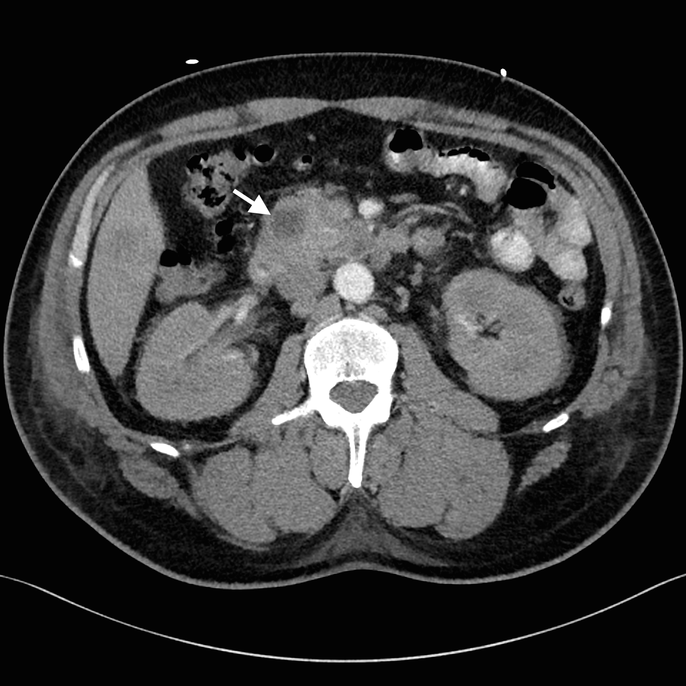
Despite adequate fluid replacement, the patient remained oliguric and developed deteriorating uraemia, so that haemodialysis was initiated. This facilitated further investigation with contrast-enhanced triple-phase hepatic CT, which confirmed a 25 × 23 × 17 mm heterogeneous head of pancreas mass, together with multiple hypodense liver lesions (Figure). Ultrasound-guided fine-needle aspiration biopsy of a liver lesion was performed. The cytological profile and immunohistochemical characteristics of the biopsy sample were consistent with metastatic adenocarcinoma, probably of pancreatic origin.

Palliative chemotherapy was considered, but the clinical condition of the patient deteriorated after the development of a pulmonary embolus and disseminated intravascular coagulopathy. He died on Day 35. Consent for an autopsy was not given.

Discussion

The hallmark of diabetic ketoacidosis (DKA) is the triad of hyperglycaemia, ketonaemia and metabolic acidosis. The pathogenesis of DKA involves a relative insulin deficiency and an excess of counterregulatory hormones.1 Interplay between these factors results in reduced glucose utilisation and increased gluconeogenesis, together with increased lipolysis and ketogenesis.

DKA is classically associated with type 1 diabetes mellitus (DM1) but it has been increasingly recognised that it may also occur in type 2 diabetes mellitus (DM2).2 Pancreatic adenocarcinoma presenting as DKA, however, is rare; only two other cases have been reported. The first involved a 75-year-old woman with longstanding DM2 who presented with DKA; pancreatic adenocarcinoma was later diagnosed.3 The second case was in a 36-year-old woman with a history of gestational diabetes; she presented with DKA associated with a pancreatic abscess that was later found to include pancreatic adenocarcinoma.4

Our patient was an otherwise healthy man who presented with first-onset DKA that led to the diagnosis of metastatic pancreatic adenocarcinoma. Despite the absence of a history of clinical symptoms, the elevated HbA1c and C-peptide levels, together with the absence of insulin autoantibodies, suggest that our patient may have had undiagnosed DM2. Further, our patient was African American, and an elevated incidence of DKA in African Americans with DM2 has been reported. In fact, studies have found that up to half of African American and Hispanic patients who developed DKA had features of DM2, referred to as “ketosis-prone DM2”.5,6 It has been hypothesised that the association of ketosis-prone DM2 with these ethnic groups reflects a genetic susceptibility to transient reductions in insulin production.5 The ethnicity of the two previous patients with pancreatic adenocarcinoma-associated DKA was unfortunately not reported.

It has traditionally been assumed that the development of DKA in people with DM2 requires a stressful precipitating event. Recent studies, however, have found that there were no obvious triggers in up to 25% of people with DM2 who developed DKA.7 For our patient, there was no identifiable trigger apart from his pancreatic adenocarcinoma. It is possible that a pancreatic adenocarcinoma may itself have either paracrine or paraneoplastic effects that disrupt normal pancreatic endocrine function. Indeed, it has been shown that even transient insulinopaenia is sufficient to elicit DKA in ketosis-prone African American patients.8 It is not known whether this played a role in our patient, as insulin and C-peptide levels were not assayed when he was initially examined.

Finally, the diagnosis of pancreatic adenocarcinoma in our patient was somewhat fortuitous, as initial abdominal imaging was undertaken to investigate the elevation in his serum pancreatic enzyme levels. A definitive diagnosis was further delayed by the patient’s acute renal dysfunction, which had initially precluded the use of intravenous contrast. A high degree of clinical suspicion of primary pancreatic disease is thus required when patients with features of DM2 present with first-onset DKA, especially if there are no apparent clinical triggers. Abdominal imaging may be warranted in these cases to look for less obvious precipitating events.

Lessons from practice

  • Diabetic ketoacidosis (DKA) has classically been associated with type 1 diabetes but there is now increasing recognition of its occurrence in type 2 diabetes.
  • It was previously assumed that relative insulinopaenia or stressful precipitating events were required to trigger DKA. Recent studies, however, indicate that there was no obvious precipitating factor in up to 25% of DKA cases in people with type 2 diabetes.
  • DKA is more common in people from certain ethnic groups (including African Americans and Hispanics) with type 2 diabetes, termed ketosis-prone type 2 diabetes.
  • If patients with type 2 diabetes present with DKA without any apparent trigger, abdominal imaging may be warranted to look for less obvious precipitating events.

Arterial phase of triple-phase computed tomography of the patient’s liver with oral contrast agent. Representative axial section showing hypodense head of pancreas mass (arrow).

Biochemical parameters of the patient immediately after his admission to hospital

Parameter

Value

Reference interval


pH

7.21

7.36–7.44

Paco2, mmHg

33

35–45

Pao2, mmHg

96

80–100

Actual bicarbonate, mmol/L

13

22–30

Base excess, mEq/L

– 14

– 2 to +2

Lactate, mmol/L

2.3

0.5–1.6


Sodium, mmol/L

166*

135–145

Potassium, mmol/L

4.8

3.5–5.0

Chloride, mmol/L

108

97–109

Bicarbonate, mmol/L

17

24–32

Urea, mmol/L

27.9

3–8

Creatinine, µg/L

391

70–110

Calcium, mmol/L

2.52

2.10–2.60

Corrected calcium, mmol/L

2.54

2.10–2.60

Glucose, mmol/L

64.8

3.0–7.7

Ketone (point-of-care), mmol/L

7.7

< 3

Osmolality, mEq/L

480

280–300


Albumin, g/L

42

38–48

Protein, g/L

85

62–80

Alanine aminotransferase (ALT), IU/L

52

5–55

Aspartate aminotransferase (AST), IU/L

23

5–55

Alkaline phosphatase (ALP), IU/L

165

30–130

γ-Glutamyl transferase (GGT), IU/L

130

< 60

Bilirubin, µmol/L

5

< 21

Amylase, IU/L

653

20–120

Lipase, IU/L

1207

13–60


Haemoglobin, g/L

178

130–170

Platelets, × 109/L

300

150–400

White cell count, × 109/L

23.1

4–10

Neutrophils, × 109/L

18.9

2.0–7.0

Lymphocytes, × 109/L

0.9

1.0–3.0

Monocytes, × 109/L

1.4

0.2–1.0


* Serum sodium corrected for hyperglycaemia is 192 mmol/L, using the formula in Banerji, et al.9


An enhanced recovery after surgery program for hip and knee arthroplasty

Osteoarthritis is the leading cause of pain and disability among the elderly and affects 15% of the population. Despite a range of treatments for osteoarthritis (OA), joint replacement remains the main treatment option for patients in whom the disease has progressed.1

In Victoria, more than 20 000 hip and knee joint replacements are now performed each year, reflecting orthopaedic practices globally. The prevalence of OA and the need for joint replacement are likely to increase because of a combination of increasing risk factors (age, obesity) and improved surgical and anaesthetic techniques that make surgery possible for more people.1

Across health services, there is wide variation in hospital length of stay for patients receiving hip and knee replacements. This is probably independent of casemix and more reflective of varying health service practices. Surgical injury, pain, stress-induced catabolism, impaired organ function and impaired cognitive function may contribute to complications, prolonged hospitalisation, postoperative fatigue, delayed convalescence and the need for rehabilitation. Optimisation of individual care components in perioperative care (the fast-track methodology) reduces the need for prolonged hospitalisation and convalescence, and reduces morbidity, with subsequent economic savings.26 Enhanced recovery after surgery (ERAS) programs are a care package of evidence-based interventions used in a multimodal, integrated clinical care pathway to achieve improved functional outcomes and rapid recovery.6 ERAS pathways have led to reduced hospital stays after hip or knee replacements — as short as 3 days in many centres.58

We aimed to assess the extent to which a predefined ERAS program for orthopaedic surgical patients could be achieved, and to evaluate improvements in quality of care and patient outcome across three public hospitals in Victoria.

Methods

We used a before-and-after study design consisting of three phases. Public health services involved in the study were the Alfred, Bendigo and Monash hospitals.

Phase 1: over the 6 months before implementation of the ERAS program, we recorded perioperative data for all eligible patients undergoing surgery (the existing-practice cohort).

Phase 2: training of staff managing orthopaedic surgical patients. For 1 month, the evidence-based background to ERAS was promulgated to all surgical, anaesthetic and nursing staff. This was done in various forms including lectures, workshops, meetings and written instructions.

Phase 3: change performance. We undertook a repeat audit following the implementation of the ERAS care package (the ERAS cohort).

Our study received ethics approval as an audit project with a waiver for specific patient consent (Alfred Human Research Ethics Committee, EC 92/12).

The pre-ERAS phase ran from March to September 2012. Training of staff took place over September 2012. The ERAS phase ran from October 2012 to May 2013.

Patient health status was quantified using the American Society of Anesthesiologists physical status classification, ranging from 1 (healthy patient) to 5 (moribund patient not expected to survive without the operation). Patient quality of recovery was assessed using the patient-centred, 15-item quality-of-recovery score,9 and the 12-item World Health Organization Disability Assessment Schedule 2.0 score.10 Indicators of patient satisfaction were assessed at 30 days after surgery using a 5-item Likert scale (0 = strongly agree, 5 = strongly disagree).

Actual hospital stay was timed from the beginning of surgery until discharge. We evaluated readiness for discharge on postoperative Day 3, defined by whether patients were eating and drinking, had no drain tubes or urinary catheters, were weight-bearing, and had well controlled pain scores (visual analogue scale with a range of 0 to 10) at rest and on movement of less than 3 and 5, respectively.

A successful ERAS implementation required at least 11 of 16 prespecified ERAS items (Box 1).

On completion of our study, there was a concern raised by the surgical team at the lead institution regarding an apparent increased incidence of acute kidney injury (AKI). In view of the widespread use of local anaesthetic infiltration with a solution that included ketorolac 30 mg, we chose to investigate this more formally at the lead institution. We retrospectively retrieved all perioperative creatinine data for the study cohorts. AKI was defined according to AKI Network11 and RIFLE (risk, injury, failure, loss, end-stage kidney disease)12 criteria. We did not include urine output or oliguria in the definitions of AKI, in part because we did not collect these data, but primarily because urine output is an unreliable indicator of renal function in the perioperative setting.

To account for the restrictive intravenous (IV) fluid regimen used in the ERAS cohort (which may have artificially elevated serum creatinine because of the avoidance of a dilutional effect from excessive IV fluids increasing body water), we calculated the adjusted creatinine concentration by first estimating the volume of distribution for creatinine as equal to total body water (assumed to be 60% of body weight, expressed in mL), and assuming that 50% of IV fluid was still accumulated as tissue oedema at the time of postoperative creatinine measurements:13

adjusted creatinine concentration = serum creatinine concentration × (1 + [0.5 × IV fluid balance/total body water])

Analysis of the data showed that there was no increased incidence of AKI in the ERAS group (Appendix 1).

The primary end point of the study was duration of hospital stay. Secondary end points were adherence to the ERAS bundle (defined as ≥ 11 items), and a number of patient outcome measures. A sample size calculation based on a change in hospital stay from a mean of 7 days (SD, 4 days) to 6 days (SD, 3 days), with an α value of 0.05 and a β value of 0.2, required at least 380 patients to be enrolled, but we included a larger sample in view of the planned subgroup analyses. Continuous data are reported as mean (SD) or median and interquartile range (IQR). Numerical data were first tested for normality using the Kolmogorov–Smirnov test and then compared using the Student t test or Wilcoxon rank–sum test, as appropriate. Rates were compared using χ2 or Fisher exact test, as appropriate. Hospital stay was expected to be skewed to the right because of a small proportion experiencing complications and a protracted hospital stay. Therefore, we log-transformed hospital stay data to enable valid comparison using the t test; in addition, we report median (IQR) length of stay and results of Wilcoxon rank–sum testing. Patients undergoing each type of surgery were also analysed as subgroups. A P value of less than 0.05 was considered statistically significant. Data were analysed with SPSS version 20.0 for Windows (SPSS Inc).

Results

We enrolled 709 patients into the project; 412 in the existing-practice cohort and 297 in the ERAS cohort (Box 2). We achieved 100% patient follow-up to hospital discharge and 90% follow-up at 6 weeks; there were 41 (10%) and 25 (8%) patients missing in each cohort, respectively. Comparison of data from the three hospitals showed that the patients were similar demographically as well as having similar rates of physical functioning and comorbidity. The existing-practice and ERAS cohorts, with the exception of some medications, were comparable (Box 2), which allowed unadjusted analyses between groups.

The ERAS program led to a significantly higher rate of successful implementation of this clinical pathway (2% v 81%; P < 0.001) (Box 3, Box 4, Box 5 and Appendix 2). The post-implementation cohort had a significantly increased number of ERAS interventions compared with the existing-practice group (median, 12 [IQR, 10–13] v 8 [IQR, 7–10]; P < 0.001).

Overall, there was a significant reduction in hospital stay (geometric mean, 5.3 [SD, 1.6] v 4.9 [SD, 1.6] days [P < 0.001]) (Box 4), with around half of the patients being discharged from hospital within 5 days of surgery (ERAS group, 60% v existing-practice group, 52%; P = 0.086). For those undergoing knee replacement surgery, the ERAS program was associated with a reduced hospital stay (geometric mean, 5.3 [SD, 1.6] v 4.5 [SD, 1.5] days [P = 0.001]; median, 5.0 [IQR, 4.0–6.7] v 4.1 [IQR, 3.0–6.0] days [P = 0.005]); and a greater proportion of patients were more likely to be discharged by Day 5 (64% v 52%; P = 0.019). There was no change in median hospital stay for hip replacement patients in the ERAS group compared with the existing-practice group (median, 5.0 [IQR, 3.5–7.0] v 5.0 [IQR, 4.0–6.9] days; P = 0.99). Overall, the 75th centile for length of stay decreased from 6.8 to 6.0 days.

We found high rates of compliance with nearly all ERAS items (Box 3). There was increased use of spinal anaesthesia. The use of femoral nerve block (with or without a catheter) was substituted by favouring surgeon-delivered local anaesthetic infiltration in 75% of cases; this change in practice varied across the three hospitals (98%, 37% and 98%). There were improved dynamic pain scores and quality of recovery (Box 4). There were improvements in other recovery parameters (early feeding, ambulation and removal of tubes). Patients undergoing knee replacement had improved flexion on postoperative Days 1 and 2.

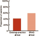
The proportion of patients ready for discharge on Day 3 after surgery was significantly higher in the ERAS group compared with the existing-practice group: 59% v 41%, respectively; relative risk, 1.35 (95% CI, 1.18–1.53); P < 0.001 (Box 6 and Appendix 1).

The 6-week complication rates were similar and there was no increase in the rate of hospital readmission. Pain levels were similar and there was a higher level of patient satisfaction at 6 weeks after surgery (Box 5).

The incidence of AKI was comparable between groups (Appendix 1). The final plasma creatinine values were slightly higher in the ERAS group, but this could be accounted for by higher baseline (preoperative) values; ERAS patients had a mean change in creatinine from 78 mmol/L (SD, 24 mmol/L) preoperatively, to a final reading of 79 mmol/L (SD, 24 mmol/L) postoperatively. There were no cases of renal failure.

Discussion

Our study results indicate that implementing an ERAS program may be beneficial for other Victorian public hospitals. The ERAS program had a small but significant effect on hospital stay, particularly for knee replacement patients. A pertinent finding from our study was that a higher proportion of patients managed through the ERAS care pathway compared with the existing-practice group (59% v 41%, respectively) were deemed ready for discharge on postoperative Day 3.

The limited effect on actual hospital stay in this project is likely to be due to one key factor: despite an effectively implemented ERAS program, there were entrenched hospital practices that prevented earlier hospital discharge even though patients were deemed ready for discharge; that is, discharge planning practice was mostly unchanged. This is due to established ward practices, including a repetitive requirement for many joint replacement patients to undergo their initial rehabilitation program as an inpatient (delaying their discharge). Patients referred for rehabilitation often wait for some time (hours or days) before being reviewed by rehabilitation services. Further, if surgery occurred on a Thursday or Friday, patients had minimal access to physiotherapy over the weekend. These aspects offer opportunities for improvement. The challenge is clear: to convert improvement in care (and outcome) into shorter hospital stay.

Our primary end point was duration of hospital stay. As we have done previously,14 we used a log-transformation and compared geometric means to account for skewness of our data (a small proportion of patients staying in hospital for very long times distorts central tendency — a well known phenomenon for many types of surgery). A secondary non-parametric comparison of median stays was not statistically significant. Therefore, we reported the 75th centiles to illustrate the observed improvement in hospital stay for the majority of patients.

Administrative and traditional patterns of clinical practice limit opportunities for change and are common causes of delayed discharge from hospital.1416 Perhaps specific fast-track arthroplasty units that have evidence-based and protocolised rapid recovery pathways can optimise cost-efficient quality outcomes after hip and knee replacement surgery.15 This could reduce hospital costs, improve patient satisfaction with care and potentially reduce perioperative morbidity.

We demonstrated that an ERAS program for orthopaedic joint replacement can be achieved. We markedly improved most indicators of processes related to an ERAS program. These included preadmission patient education, reduced fasting times, clear oral fluids, written instructions (including expected day of discharge), less blood loss, better pain relief, earlier ambulation and better overall quality of recovery. Similar success has been reported in other countries.8,17,18 Medical teams can be trained to deliver an ERAS program and this clearly improves the quality of care.

We clearly demonstrated that we could successfully implement a predefined ERAS program for orthopaedic surgical patients in public hospitals, and that doctors and nurses could follow such a regimen to improve outcome parameters. There was high uptake of nearly all ERAS items. This led to clinically important improvements in care, a small reduction in hospital stay for knee replacement patients, and an overall improvement in some aspects of patient satisfaction. There was evidence of improved quality of recovery. There was no effect on most complications and no adverse effect on hospital readmission rates.

Our 6-week complication rates are lower than those reported in most studies, the largest of which included 4500 consecutive unselected hip and knee replacements.5 In that study, the first 3000 patients represented existing or traditional practice and a further 1500 patients underwent an ERAS protocol similar to that used in our study. This group reported a decrease in length of stay from 6 days to 3 days (P < 0.001), as well as a reduction in 30-day mortality (from 0.5% to 0.1%; P = 0.02) and 90-day mortality (from 0.8% to 0.2%; P = 0.01). Blood transfusion was reduced from 23% to 9.8% (P < 0.001). There was a trend of a reduced rate of 30-day myocardial infarction (from 0.8% to 0.5%; P = 0.2) and stroke (from 0.5% to 0.2%; P = 0.2). There was no measurable effect on deep vein thrombosis (0.8% v 0.6%; P = 0.5) or pulmonary embolism (1.2% v 1.1%; P = 0.9).

There were some limitations of our study. It was not a randomised trial and there may have been some imbalance between the groups that we did not account for. The study was unblinded and we compared numerous secondary end points. Although there are likely to be cost benefits of an ERAS program, we did not undertake any health costing analyses.

We found a high level of general acceptance and uptake of the ERAS program and, on the whole, it had a positive effect on patients and staff. We can recommend this orthopaedic ERAS pathway.

Anchor

1 Sixteen predefined enhanced recovery after surgery items for hip or knee arthroplasty

  • Nurse coordinator counselling in the orthopaedic or preadmission clinic
  • Preadmission review by a physiotherapist and/or dietitian
  • Minimal fasting preoperatively, defined as clear oral fluids up to 2 hours before surgery
  • Preoperative oral carbohydrate loading
  • No sedative premedication (benzodiazepines, opioids or neuroleptics)
  • Pre-emptive analgesia with paracetamol and gabapentinoids according to protocols
  • Spinal anaesthesia (not epidural)
  • Local anaesthesia technique (surgeon-delivered local infiltration of analgesia or anaesthetic femoral nerve block)
  • Minimal (≤ 10 mg) intravenous morphine intraoperatively
  • Intraoperative avoidance of excessive intravenous fluids (knee, > 1 L; hip, > 2 L; both: subtracting blood loss)
  • Active intraoperative warming (forced air warming and/or warmed intravenous fluids)
  • Antiemetic prophylaxis
  • Multimodal oral analgesia for ≥ 3 days postoperatively, to include a non-steroidal anti-inflammatory drug or cyclooxygenase-2 inhibitor
  • Early postoperative (recovery room) oral carbohydrate supplementation
  • Mobilisation within 24 hours
  • Early hospital discharge (≤ 5 days)

2 Patient demographic and perioperative characteristics*

Variable

Existing practice (n = 412)

ERAS (n = 297)

P


Sex, male

164 (40%)

113 (38%)

0.64

Mean age, years (SD)

68 (11)

67 (10)

0.22

Mean weight, kg (SD)

84 (19)

87 (20)

0.092

Medical history


Current smoker

46 (11%)

30 (10%)

0.65

Hypertension

284 (69%)

194 (65%)

0.31

Coronary artery disease

70 (17%)

41 (14%)

0.25

Stroke

20 (5%)

10 (3%)

0.33

Heart failure

19 (5%)

15 (5%)

0.86

Peripheral vascular disease

13 (3%)

8 (3%)

0.72

Diabetes

81 (20%)

73 (25%)

0.12

COPD

88 (21%)

67 (23%)

0.70

Preoperative anaemia

36 (9%)

22 (7%)

0.52

Usual medications


Opioid

106 (26%)

58 (20%)

0.053

Aspirin within 5 days

58 (14%)

54 (18%)

0.14

Clopidogrel within 7 days

2 (< 1%)

0

0.23

Warfarin within 7 days

22 (5%)

6 (2%)

0.025

NSAID/COX-2 inhibitor

109 (26%)

109 (37%)

0.004

ACE inhibitor/ARB

235 (57%)

168 (57%)

0.90

Beta blocker

71 (17%)

51 (17%)

0.98

Statin

141 (34%)

89 (30%)

0.37

Calcium channel blocker

98 (24%)

73 (25%)

0.81

Diuretic

120 (29%)

79 (27%)

0.45

Oral hypoglycaemic

57 (14%)

60 (20%)

0.024

Insulin

9 (2%)

11 (4%)

0.25

LMWH

20 (5%)

8 (3%)

0.15

ASA physical status


0.57

1

10 (2%) 10 (3%)  

2

223 (54%) 161 (54%)  

3

172 (42%) 122 (41%)  

4

7 (2%)

3 (1%)


Disease

    0.22

Osteoarthritis

367 (89%) 275 (93%)  

Rheumatoid arthritis

4 (1%)    

Avascular necrosis

14 (3%) 7 (2%)  

Other

27 (7%) 15 (5%)  

Previous PONV or motion sickness

124 (30%) 81 (27%) 0.41

ACE = angiotensin-converting enzyme. ARB = angiotensin-receptor blocker. ASA = American Society of Anesthesiologists. COPD = chronic obstructive pulmonary disease. COX = cyclooxygenase. ERAS = enhanced recovery after surgery, LMWH = low molecular weight heparin. NSAID = non-steroidal anti-inflammatory drug. PONV = postoperative nausea and vomiting. * Data are no. (%) of patients unless otherwise specified. † P value derived from χ2 test for trend.

3 Perioperative and surgical care*

Variable

Existing practice (n = 412)

ERAS (n = 297)

P


Preadmission clinic, seen by:


Nurse

406 (99%)

297 (100%)

0.037

Anaesthetist

405 (98%)

297 (100%)

0.024

Surgeon

406 (99%)

297 (100%)

0.037

Physiotherapist

331 (80%)

224 (75%)

0.12

Occupational therapist

324 (79%)

249 (84%)

0.077

Dietitian

0

61 (21%)

< 0.001

Day of surgery


Admission on day of surgery

404 (98%)

294 (99%)

0.32

Shower with antibiotic soap

399 (97%)

238 (80%)

< 0.001

Preoperative skin wipes

4 (1%)

91 (31%)

< 0.001

Oral (clear) fluids given

2 (< 1%)

180 (61%)

< 0.001

Oral carbohydrate drink

0

248 (84%)

< 0.001

Gabapentin premedication

21 (5%)

172 (58%)

< 0.001

Type of surgery


Hip

214 (52%)

129 (43%)

0.025

Knee

198 (48%)

168 (57%)

0.025

Revision

26 (6%)

13 (4%)

0.26

Tubes


Urinary catheter

200 (49%)

107 (36%)

0.001

Drain tube(s)

105 (25%)

59 (20%)

0.080

Type of anaesthesia


General (± regional)

266 (65%)

164 (55%)

0.014

Spinal

236 (57%)

205 (69%)

0.001

Epidural or CSE

16 (4%)

3 (1%)

0.019

Postoperative regional analgesia


Nerve block used

135 (33%)

44 (15%)

< 0.001

LA infiltration

214 (52%)

222 (75%)

< 0.001

PONV prophylaxis

233 (57%)

202 (68%)

0.002

Mean total IV fluids, mL (SD)

1756 (767)

1446 (687)

< 0.001

Active (forced air) warming

392 (95%)

285 (96%)

0.34

Mean lowest temperature, °C (SD)

35.7 (0.5)

36.2 (0.4)

0.039

Mean duration of surgery, hours (SD)

2.0 (0.9)

1.9 (0.6)

0.33


CSE = combined spinal and epidural. ERAS = enhanced recovery after surgery. IV = intravenous. LA = local anaesthesia. PONV = postoperative nausea and vomiting. * Data are no. (%) of patients unless otherwise specified. † Key ERAS implementation points.

4 Recovery profile and hospital stay

Variable

Existing practice (n = 412)

ERAS (n = 297)

P


Recovery room


Median pain score (IQR), at rest*

0 (0–5)

0 (0–4)

0.047

Median pain score (IQR), on movement*

0 (0–7)

0 (0–4)

< 0.001

Admission temperature, °C (SD)

36.1 (0.5)

36.0 (0.6)

0.006

Postoperative, at 24 hours


Median pain score (IQR), at rest*

5 (3–7)

4 (2–5)

< 0.001

Median pain score (IQR), on movement*

6 (4–8)

5 (3–7)

< 0.001

Mean quality of recovery score (SD) (range, 0–150)

103 (19)

106 (20)

0.056

Total knee replacement


Mean knee flexion (SD), degrees

51 (19) 57 (24) 0.026

Median quadriceps strength (IQR), Nm

3 (2–3)

2 (2–3)

0.11

Postoperative, at 48 hours


Median pain score (IQR), at rest*

4 (2–6)

3 (1–5)

< 0.001

Median pain score (IQR), on movement*

6 (4–8)

5 (2–7)

0.001

Total knee replacement


Mean knee flexion (SD), degrees

72 (19)

78 (14)

0.009

Median quadriceps strength (IQR), Nm

3 (2–3)

3 (2–3)

0.90

Recovery parameters, median hours (IQR)


Weight bearing

1.1 (1.0–2.0)

1.0 (0.9–2.0)

0.001

Oral fluid intake

3.2 (2.0–5.0)

2.7 (1.7–4.1)

0.016

Oral food intake

7.0 (4.3–15)

6.3 (3.2–7.9)

0.004

Removal of drain tubes

27 (24–42)

25 (23–27)

0.002

Removal of urinary catheter

48 (42–76)

33 (17–60)

< 0.001

Blood transfusion in hospital, no. of patients (%)

58 (14%)

31 (10%)

0.24

Return to theatre, no. of patients (%)

14 (3%)

10 (3%)

0.76

Length of stay, days


Geometric mean (SD)

5.3 (1.6)

4.9 (1.6)

< 0.001

Median (IQR)

5.0 (4.0–6.8)

5.0 (3.8–6.2)

0.10


ERAS = enhanced recovery after surgery. IQR = interquartile range. * Visual analogue scale: 0 = none, 10 = severe.

5 Recovery profile at 6 weeks after surgery*

Variable

Existing practice (n = 412)

ERAS (n = 297)

P


Wound infection

21 (5%)

13 (4%)

0.99

Prosthesis infection

5 (1%)

2 (< 1%)

0.60

Prosthetic joint dislocation

2 (< 1%)

3 (1%)

0.31

Periprosthetic fracture

0

0

> 0.99

Thromboembolism

13 (3%)

10 (3%)

0.59

Urinary tract infection

8 (2%)

2 (1%)

0.22

Death

2 (< 1%)

1 (< 1%)

0.85

Worst pain rating in past 24 hours

2 (0–4)

2 (0–3)

0.01

Extent of disability in past 24 hours

2 (0–3)

1 (0–2)

0.37

Patient satisfaction


Was surgery worthwhile?

1 (1–2)

1 (1–1)

< 0.001

Did surgery improve your daily life?

1 (1–2)

1 (1–2)

0.015

Do you feel better?

1 (1–2)

1 (1–2)

< 0.001

Do you have trouble sleeping?

3 (2–4)

3 (2–4)

0.67

Hospital readmission

25 (6%)

15 (5%)

0.87


ERAS = enhanced recovery after surgery. * Data are no. (%) of patients or median score (IQR). † Visual analogue scale: 0 = none, 10 = severe. ‡ 5-point Likert scale: 0 = strongly agree, 5 = strongly disagree.


6 Proportion of patients ready for discharge on Day 3 after surgery*


ERAS = enhanced recovery after surgery. * P < 0.001.

Specialist unemployment: time to be worried?

Consultants working for free to maintain their recency of practice. New Fellows accepting positions with reduced conditions to get their foot in the door. Others prompted to work part time or as locums. Young Fellows choosing to do more sub-specialty training because they cannot find work in their chosen field.

Apocryphal stories maybe, but there is genuine concern in the profession that some specialists are experiencing underemployment or even unemployment.

“Exit block” from training – where recently graduated Fellows stay in training positions that would otherwise be filled by specialist trainees because the consultant jobs aren’t there − is a knock-on effect from this scenario.

We should be worried about shrinking employment opportunities for new Fellows and exit block for specialist trainees.

Among other things, it would mean that some specialists are struggling to get the workload they need to keep their skills fresh. Nor would trainees be getting access to the positions that provide the clinical cases they require to complete their specialist training. It would ultimately mean that Australia is squandering its considerable investment in the medical workforce over the past decade.

So are we really seeing the early signs of an oversupply of specialists, or is the issue a poorly distributed workforce?

Unfortunately, there is no hard data, but anecdotal reports of underemployment and unemployment in some specialties are emerging.

A specialty that might be affected is anaesthesia, where there is increasing concern that an oversupply of anaesthetists is looming. There is a range of possible reasons for this situation, including the large numbers of anaesthesia trainees employed by public hospitals; fewer opportunities for consultants in the public system; fewer private sector opportunities in major metropolitan areas; difficulties in getting credentialing at private hospitals; and senior specialists delaying their retirement.

I met with the Australian and New Zealand College of Anaesthetists and the Australian Society of Anaesthetists in January to discuss the state of the anaesthesia workforce.

Surveys of new Fellows run by both organisations showed that some had experienced unemployment and underemployment after gaining Fellowship, and were concerned about future career prospects.

I understand that the situation in anaesthesia could be emerging in some other specialties as well.

The outcome of the meeting was a joint submission to the National Medical Training Advisory Network (NMTAN) asking it to include the anaesthetist workforce in its modelling program as a matter of urgency. Pleasingly, it has told us that this is indeed a priority for the network.

The AMA is being proactive in getting an understanding of the scale of specialist unemployment across the specialties. The Medical Workforce Committee is taking the lead, working closely with our doctors in training.

We need to get this right and find out whether an oversupply of specialists is building, or whether the problem is one of distribution.

Both scenarios would have obvious, and very different, implications for developing and coordinating the future medical workforce.

Separate to the joint submission on the anaesthetist workforce, the AMA has asked NMTAN to undertake the data collection needed to determine what’s happening across all specialties, and identify the measures needed to ensure that, subject to community demand for medical services, there will be sufficient jobs for doctors when they finish their training.

I’m hopeful that NMTAN is taking the issue seriously.

In the meantime, we are liaising with the Colleges on how their new Fellows are faring.

While we don’t want to generate unnecessary angst among trainees on their job prospects, it is important that they have a clear idea of future workforce scenarios when they make their career choices.

Effectiveness of a care bundle to reduce central line-associated bloodstream infections

Central line-associated bloodstream infections (CLABSIs) are an important source of morbidity, mortality and cost.1 About 4000 CLABSIs occur in Australian intensive care units (ICUs) each year, with an estimated nationwide cost of $36.26 million and a mortality rate of 4%–20%.2,3 The importance placed on CLABSI and its prevention has prompted standardised monitoring for quality assurance and innovation of preventive strategies.1,4,5 Care bundles focused on improving line insertion procedure have proven successful overseas.1,6 Local implementation of a similar care bundle to that used overseas across New South Wales proved successful, and prompted the Australian and New Zealand Intensive Care Society CLABSI Prevention Project.7,8 Despite these interventions, CLABSI rates range from 0.9 to 3.6 per 1000 central line days.6,7,920

The Victorian Healthcare Associated Infection Surveillance System (VICNISS) collects standardised ICU CLABSI rates for the state of Victoria.21 Since 2006, the University Hospital Geelong (UHG) ICU has reported CLABSI rates to VICNISS.

An elevated reported CLABSI rate at UHG in 2007 and 2008 (3.8 and 3.6, respectively, compared with the state average of 2.7 per 1000 central line days)22 prompted development and introduction of a CLABSI prevention care bundle. Our care bundle used an effective line insertion procedure identified from previous studies,1,6,7 but also incorporated a novel maintenance procedure. In this article, we report the effectiveness of this care bundle in a tertiary ICU in Victoria.

Methods

We undertook a before-and-after study, retrospectively accessing the pre-intervention data, at an adult, tertiary, 19-bed ICU that admits medical, surgical and cardiac surgical patients. Ethics approval was obtained from the Barwon Health Research Review Committee. This project was performed as part of the authors’ usual roles and no funding or subsidy was received. All of us had full access to the study data.

Intervention

The care bundle was based on the Australian and New Zealand Intensive Care Society CLABSI prevention project,8 comprehensive literature review and collaboration between UHG ICU, UHG Infection Control Services and other key stakeholders. The final care bundle (Appendix 1) included standard line insertion procedure consistent with that described previously,6,7 bedside audit by an observer with stopping rules, and a novel line maintenance procedure that included placement of a Biopatch (Johnson and Johnson), sterile line access, daily 2% chlorhexidine body wash, daily central venous catheter (CVC) review with early line removal, and liaison nurse follow-up of all CVCs present at discharge.

Study procedure

All adult patients admitted to UHG ICU between 1 July 2006 and 30 June 2014 were captured in this study. The care bundle was introduced in 2009, dividing patients into a pre-intervention period (1 July 2006 to 31 December 2009) and a post-intervention period (1 January 2010 to 30 June 2014). Case identification of CLABSI was based on the VICNISS dataset and review of blood cultures. All VICNISS-reported CLABSI cases were reviewed by one of us (D E) to confirm that they fulfilled the current VICNISS definition (Appendix 2). This definition is consistent with the internationally accepted O’Grady definition that has been previously applied.7,23

All confirmed CLABSIs were included in the analysis, irrespective of whether line insertion occurred in the ICU. Cohort demographic, basic clinical and microbiological data were collected from the hospital electronic database. Patient medical records of all VICNISS-reported CLABSI cases were reviewed to confirm CLABSI definition and collect additional clinical information. Finally, all positive blood cultures were blindly and independently reviewed by an infectious diseases specialist to identify any missing CLABSI cases.

Statistical analysis

Data were analysed using SAS, version 9.4 (SAS Institute). All data were visually assessed for normality using histograms. The primary outcome (CLABSI events) was compared first as an overall comparison of proportions and presented as a relative risk with 95% confidence intervals and second as the number of CLABSI events per quarter using Poisson regression.

Comparisons of pre- and post-intervention periods were performed for categorical variables using χ2 tests for equal proportions and reported as numbers (%). Normally distributed variables were compared using Student t tests and reported as mean (SD), and non-normally distributed data were compared using Wilcoxon rank-sum tests and reported as median (interquartile range). A two-sided P of 0.05 was considered to be statistically significant.

Results

Patient cohort characteristics are detailed in Box 1. The post-intervention cohort was significantly younger (mean age, 59.4 years v 64.2 years; P < 0.001) with a higher mean illness severity score (Acute Physiology and Chronic Health Evaluation [APACHE] III score, 50 v 48; P = 0.001), an increased proportion of medical patients (3250/6273 [52%] v 1863/4701 [40%]; P < 0.001), an increased requirement for mechanical ventilation (3223/6273 [51%] v 2014/4701 [43%]; P < 0.001) and an increased admission source from the wards or emergency department. Although the clinical significance of the differences in age and APACHE score are questionable, when all differences are considered together, they favour an increased risk of CLABSI in the post-intervention cohort.

A total of 24 783 central line days occurred between July 2006 and June 2014 (Box 2). Thirty cases of CLABSI were included in the analysis (eight did not satisfy CLABSI definition criteria and were excluded — seven pre-intervention and one post-intervention; Appendix 3). No CLABSI cases additional to VICNISS-reported cases were identified. In the pre-intervention period, there were 9844 central line days and 22 cases of CLABSI, resulting in a CLABSI rate of 2.2/1000 central line days. In the post-intervention period, there were 14 939 central line days and eight cases of CLABSI, resulting in a CLABSI rate of 0.5/1000 central line days. This represents a rate ratio of 0.23 (95% CI, 0.11–0.54; P = 0.005). The temporal change in CLABSI rates is shown in Appendix 3, with a peak CLABSI rate of 5.2/1000 (4/766) central line days in quarter 4 of 2008, and a CLABSI rate of zero since June 2012. The difference in the quarterly CLABSI rate before and after the intervention was introduced was significant (P < 0.001), as was the difference in the number of quarters in which CLABSI rate was zero (pre-intervention, 3/14 v post-intervention, 12/18; P = 0.01).

The blood culture collection rate (60.1 [2827/4701] v 61.5 [3859/6273] per 100 patients) was similar in the pre- and post-intervention periods, while the positive culture rate significantly fell from 9.1% (258/2827) to 7.2% (279/3859) (P = 0.005) (Box 2). Characteristics of the confirmed CLABSI cases are presented in Box 3. The site of blood culture collection was similar between the two cohorts; however, no common skin commensals were isolated as a causative organism in the post-intervention cohort.

Discussion

Our study describes a significant reduction in the CLABSI rate in a tertiary Australian Victorian ICU from a peak quarterly rate of 5.2 to zero after implementation of a care bundle that incorporated a novel line maintenance procedure. Overall, the CLABSI rate, per 1000 central line days, decreased from 2.2 in the pre-intervention period to 0.5 in the post-intervention period. In real terms, the reduced CLABSI rate equates to 15 fewer cases of CLABSI for the post-intervention period with an estimated total reduction in ICU length of stay of 38 days, hospital length of stay of 113 days and resultant cost saving of about $210 000.

To our knowledge, this is the first time that a zero CLABSI rate has been achieved and sustained in an Australian ICU. Burrell and colleagues reported a CLABSI rate of 0.9/1000 central line days from several centres.7 Department of Health data from Western Australia have shown similarly low CLABSI rates, but their processes were not reported.5,7,24

The finding of clinical effectiveness after introduction of the care bundle suggests that the observed benefits are causally associated. It is plausible that the maintenance procedure was crucial in reducing CLABSI, given that zero CLABSI was achieved despite the inclusion of lines inserted outside the ICU. It remains possible that changes in the patient cohort or procedures relating to CLABSI surveillance could account for the observed changes. In particular, there were seven CLABSIs that did not meet definition criteria in the pre-intervention period compared with one in the post-intervention period, raising the possibility of previous overreporting. Otherwise, the identified post-intervention cohort changes when taken together are considered as predisposing to CLABSI. In addition, the central line days and blood cultures per patient do not support altered clinical practice as an explanation.

Our study’s strengths include a large patient cohort with availability of population characteristics, a microbiological blood culture dataset, an independent review of all positive blood cultures, and the application of the current standard CLABSI definition across the entire study period. This reduces the likelihood that the observed change was driven by changes in non-infection control related clinical practices. This study is limited by a single-centre retrospective, observational design, limiting generalisability and the ability to establish causality. However, these limitations are largely comparable to prior similar studies.6,7,25 Other limitations included potential confounding from lines inserted outside the ICU and the absence of adherence data for the individual components of our line maintenance procedure to show actual change in clinical practice. However, in our experience, the care bundle has been embedded into routine and has markedly improved clinical practice.

In conclusion, our study suggests that a central line care bundle with this novel line maintenance procedure can effectively reduce the CLABSI rate to zero and that this zero CLABSI rate can be sustained. Validation of our study by other centres, especially if performed prospectively, would further support our findings.

1 Patient population characteristics, and ICU interventions and outcomes, for the pre- and post-intervention periods


Pre-intervention

Post-intervention

P


No.

4701

6273


Mean age in years (SD)

64.2 (16.6)

59.4 (21.2)

< 0.001

Male, no. (%)

2870 (61%)

3857 (61%)

0.64

Median APACHE III score (IQR)

48 (37–64)

50 (38–67)

0.001

Comorbidity, no. (%)


Respiratory

233 (5%)

204 (3%)

< 0.001

Cardiovascular

453 (10%)

176 (3%)

< 0.001

Hepatic

42 (1%)

117 (2%)

< 0.001

Renal

103 (2%)

134 (2%)

0.84

Immunosuppression

271 (6%)

544 (9%)

< 0.001

Cancer

231 (5%)

301 (5%)

0.78

Category, no. (%)


Medical

1863 (40%)

3250 (52%)

< 0.001

Surgical

1071 (23%)

1027 (16%)

< 0.001

Cardiac surgical

1767 (38%)

1996 (32%)

< 0.001

ICU admission source, no. (%)


Operating theatre

2752 (59%)

2988 (48%)

< 0.001

Emergency department

910 (19%)

1565 (25%)

< 0.001

Ward

801 (17%)

1280 (20%)

< 0.001

Other ICU

235 (5%)

439 (7%)

< 0.001

ICU outcomes


Mechanical ventilation, no. (%)

2014 (43%)

3223 (51%)

< 0.001

Median ICU stay in hours (IQR)

41.2 (22.3–65.9)

41.7 (21.7–73.3)

0.01

Median hospital stay in days (IQR)

9.9 (5.8–18.9)

9.0 (5.1–16.6)

< 0.001

ICU mortality, no. (%)

333 (7%)

423 (7%)

0.70

Hospital mortality, no. (%)

534 (11%)

672 (11%)

0.34


APACHE = Acute Physiology and Chronic Health Evaluation. ICU = intensive care unit. IQR = interquartile range.


2 Summary of total ICU patient admission, central line, blood culture and CLABSI data for the pre- and post-intervention periods


Pre-intervention

Post-intervention

Rate ratio (95% CI)

P


Total patient days

8070

10 899


Total central line days

9844

14 939


Central line days per patient days

1.22

1.37


Total blood cultures

2827

3859


Blood cultures per patient days

0.35

0.36

1.01 (0.97–1.05)

0.59

ICU bacteraemia, no. (%)

258 (9.1%)

279 (7.2%)

0.79 (0.67–0.93)

0.005

CLABSI, no.

22

8

0.23 (0.11–0.54)

0.005

CLABSI rate per 1000 central line days

2.2

0.5


CLABSI = central line-associated bloodstream infection. ICU = intensive care unit.


3 Characteristics of CLABSI cases for the pre- and post-intervention periods

Characteristics of infected lines

Pre-intervention (n = 22)

Post-intervention (n = 8)


Line type, no. (%)


CVC

21 (95%)

6 (75%)

Vascath

7 (32%)

3 (38%)

PAC

1 (5%)

1 (13%)

Other

3 (14%)

2 (25%)

Median dwell time, days (IQR)

6 (5–8)

5 (4–6)

Inserted in ICU, no. (%)

15 (68%)

7 (88%)

CLABSI organism, no. (%)

Staphylococcus aureus

6 (27%)

2 (25%)

Staphylococcus epidermidis

6 (27%)

0

Enterobacter spp.

1 (5%)

2 (25%)

Candida spp.

6 (27%)

2 (25%)

Enterococcus

4 (18%)

1 (13%)

Other

2 (9%)

3 (38%)

Positive blood culture site, no. (%)

Peripheral

5 (23%)

2 (25%)

Arterial

1 (5%)

0

Central

8 (36%)

2 (25%)

Unknown

19 (86%)

5 (63%)


CLABSI = central line-associated bloodstream infection. CVC = central venous catheter. ICU = intensive care unit. IQR = interquartile range. PAC = pulmonary artery catheter.


Local acquisition and nosocomial transmission of Klebsiella pneumoniae harbouring the blaNDM-1 gene in Australia

The emergence of carbapenem-resistant Enterobacteriaceae constitutes a critical global issue. Isolates harbouring the metallo-β-lactamase gene blaNDM-1 have few available treatment options. We report a case of an Australian adult with a locally acquired, community-onset blaNDM-1 Klebsiella pneumoniae infection and likely nosocomial transmission to another patient.

Clinical records

Patient A, a 68-year-old Australian-born woman living with her husband and son, had never travelled overseas and had no known contact with overseas visitors. Her past history included chronic bilateral lymphoedema with recurrent lower limb cellulitis, requiring multiple previous hospital admissions and home nursing care. She presented with septic shock and right leg erythema surrounding a 10 × 10 cm ulcer near the right lateral malleolus. Magnetic resonance imaging showed bony oedema and enhancement in the lateral malleolus, suggestive of osteomyelitis. Pseudomonas aeruginosa was isolated from blood cultures. A tissue biopsy from the overlying ulcer cultured P. aeruginosa, non-multiresistant methicillin-resistant Staphylococcus aureus (NORSA), and carbapenem-resistant Klebsiella pneumoniae, resistant to all first-line antimicrobials tested and susceptible to only colistin and fosfomycin.

She was given intravenous ceftazidime and vancomycin for treatment of P. aeruginosa and NORSA infection. After 6 weeks of treatment, the ulcer was not healing, and treatment for the carbapenemase-producing K. pneumoniae was commenced with intravenous colistin methanosulfonate (120 mg colistin base activity 12-hourly). Colistin was ceased after 3 weeks owing to acute kidney injury, and oral fosfomycin (3 g every 3 days) was administered for a further 6 weeks, in addition to oral ciprofloxacin and rifampicin for ongoing treatment of NORSA and P. aeruginosa infection. Since the cessation of her antibiotics, she has not required further antibiotic treatment of her ulcers.

Patient B, a 35-year-old Australian-born man with no history of overseas travel, was admitted with bilateral thigh cellulitis and septic shock. He had bilateral thigh debridement, with no evidence of necrotising fasciitis. Methicillin-susceptible S. aureus (MSSA) was isolated from multiple blood cultures. MSSA and Serratia liquefaciens were isolated from a thigh wound swab culture. Transthoracic echocardiogram revealed a left ventricular thrombus. He was commenced on intravenous flucloxacillin and ciprofloxacin. Two weeks after admission, further surgical samples from his thigh wounds cultured carbapenem-resistant K. pneumoniae, with similar antimicrobial susceptibility phenotype to Patient A. The isolate was deemed to be colonising the wound only, and no antimicrobials were commenced for treatment.

Both K. pneumoniae isolates demonstrated carbapenemase activity using the Carba NP assay.1 Molecular tests using polymerase chain reaction and sequencing for carbapenemase, extended-spectrum β-lactamase, plasmid-mediated AmpC β-lactamase and 16S ribosomal RNA methylase genes were performed as described elsewhere.2 The metallo-β-lactamase gene blaNDM-1 was detected in both isolates, in addition to blaCTX-M-15 and 16S ribosomal RNA methylases (armA and rmtB), which confer resistance to aminoglycosides, including amikacin. The relatedness of isolates was determined by semi-automated repetitive sequence-based polymerase chain reaction using a DiversiLab Klebsiella kit (bioMérieux). This analysis showed a > 95% genetic similarity between the two isolates. Further, the isolates were genetically distinct from two blaNDM-1-harbouring isolates that we isolated previously in patients with a history of overseas travel.

Patients A and B were admitted in December 2013 to a high dependency unit (HDU) — a four-bed area separated by curtains. They were one bed apart for 5 days before being moved adjacent to each other for 1 day, with Patient B occupying the bed cubicle space formerly occupied by Patient A for a further 5 days. The carbapenem-resistant K. pneumoniae was first identified in Patient A in the HDU, and 2 weeks later was isolated from Patient B.

After detection of the carbapenem-resistant K. pneumoniae, strict contact precautions were implemented. Environmental cleaning of the four-bed HDU and other rooms occupied by Patients A and B was undertaken with microfibre and steam cleaning, which has been shown to be an effective cleaning method.3 An exposure investigation was conducted, with environmental sampling and screening rectal swabs collected from all direct patient contacts of Patients A and B inoculated on chromogenic selective media. No other carbapenem-resistant organisms containing blaNDM-1 were isolated from clinical, screening or environmental samples.

Discussion

The metallo-β-lactamase gene blaNDM-1 was first described in a patient hospitalised in Sweden after travel to India in 20084 and subsequently identified in a series of patients in the United Kingdom, many of whom had travelled to the Indian subcontinent.5 There have been documented cases of infection with imported blaNDM-1-containing bacteria in Australia.610 These carbapenem-resistant bacteria are challenging to treat, as available treatment options are often limited to infrequently used drugs such as colistin, fosfomycin and tigecycline.

The case of Patient A has significant public health ramifications as the first detection of carbapenem-resistant blaNDM-1-harbouring K. pneumoniae infection locally acquired in Australia, independent of international travel or documented contact with a traveller. The case suggests that there may be more cases of blaNDM-1-harbouring bacteria in our community than previously suspected. We hypothesise that this carbapenem-resistant Enterobacteriaceae isolate was acquired after transmission from an unidentified carrier of blaNDM-1, possibly during previous hospital admissions or receipt of home nursing care.

The transmission from Patient A to Patient B may have occurred via a number of mechanisms. First, environmental contamination may have contributed, given that both patients shared the same bed area at different times. We previously reported an outbreak of carbapenem-resistant Enterobacteriaceae harbouring the metallo-β-lactamase gene blaIMP-4, associated with contaminated sinks in an intensive care unit; in that case, no carbapenem-resistant organisms containing blaNDM-1 were isolated from environmental samples.11 The second possible contributing factor for transmission is lapses in infection control practices by health care staff, which emphasises the importance of adhering to standard precautions such as hand hygiene.12

These cases highlight the evolving Australian epidemiology of multidrug-resistant organisms, particularly bacteria harbouring blaNDM-1. Such resistance is no longer exclusively associated with obvious international travel. There is an increasing need for effective antimicrobial stewardship and infection control measures to prevent potential future nosocomial spread of these organisms. In addition, further research and surveillance is needed in monitoring these local isolates to identify potential risk factors for local acquisition and any reservoirs within the Australian health system and community.

Are potential organ donors missed on general wards? A 6-month audit of hospital deaths

In the decade to 2008, the deceased donor and organ transplant rates in Australia failed to increase in line with population growth, and there was little change in the number of patients needing organ transplantation.1 In response to this, the Australian Government set out the National Reform Programme, comprising nine measures to establish the world’s best practice in organ and tissue donation.2

An important part of the national approach is the DonateLife Audit, which aims to report on all actual and potential organ donation activity: donor identification, request and consent rates; reasons why donation does not proceed; and missed donation opportunities. Data are collected on all deaths of patients aged between 28 days and 80 years in the emergency department (ED) and intensive care unit (ICU) (or on the wards if discharged from the ED or ICU in the previous 24 hours) and deaths of any other patient when organ donation is considered.

Royal Prince Alfred Hospital has been contributing to the DonateLife Audit since its inception, and we believe that we miss very few potential organ donors from EDs and ICUs. The DonateLife Audit does not, however, consider whether potential organ donors on the general wards who have not been recently discharged from the ICU or ED have been missed.

The success of organ donation programs is defined by the rate of deceased organ donors per million population (dpmp). Australia’s rate increased from 9–12 dpmp in 2009 to over 16 dpmp in 2013.3 Despite this, there is a body of opinion in Australia that progress has been too slow and not reflective of the large increase in funding that the reform committed.4 Furthermore, the change has not been uniform, with New South Wales achieving only 14.2 dpmp in 2013.

The increased donation rate falls well short of the rates reported for the highest performing countries, such as Spain (over 35 dpmp).5 It has been suggested that not all potential donors are being identified in Australian hospitals and that changes in hospital practice are needed to further increase donation rates.4,6

We conducted an audit of hospital deaths to examine whether potential organ donors outside the DonateLife Audit areas of EDs and ICUs are being missed. The potential for tissue-only donation was not investigated.

Methods

The audit was conducted at Royal Prince Alfred Hospital, a metropolitan 700-bed tertiary referral and teaching hospital in NSW. Specialties include neurology and neurosurgery, patients include rural and out-of-catchment referrals and patients admitted through the ED, and there is a 50-bed intensive care floor. Hospital deaths between 1 July and 31 December 2012 were reviewed by two donation specialists medical (DSMs) (both intensive care specialists) and a donation specialist nurse (DSN).

The following groups of patients were excluded from further review as they are generally deemed unsuitable for organ donation: those who died when they were aged ≥ 80 years; those admitted to hospital under oncology, palliative care for cancer or haematology services (ie, those with an oncological diagnosis); and those who could not be resuscitated from cardiac arrest in the ED. Neonates who died when they were aged ≤ 28 days were excluded, in keeping with the DonateLife Audit.

Patients referred to the DonateLife team were categorised according to standard potential organ donor categories by the DSN (Box 1).7 The remaining patients were then assessed independently for suitability and likelihood of progression to organ donation by the two DSMs, using the hospital’s electronic medical records. Where there was disagreement, the DSN reviewed the case record and had the casting vote.

Patients were deemed not medically suitable (NMS) if they were aged > 65 years and had a non-neurological diagnosis, as such patients would have been highly unlikely to become brain dead and were over the age accepted in NSW in 2012 for donation after circulatory death (DCD). Patients who had active cancer, had septicaemia or were dying a circulatory death despite maximal medical therapy were also deemed NMS, as these conditions contraindicate organ donation. Patients who died with multiple organ failure (defined as presence of two or more organ failures) were analysed individually to establish whether non-failed organs might have been suitable for donation. Finally, patients were deemed NMS if a treatment limitation stating that they were not to receive mechanical ventilation had been made.

The remaining patient deaths, where we could not establish a clear reason to exclude the potential for organ donation, were reviewed in detail and assigned to potential organ donor categories by a panel of five organ donation specialists. The panel consisted of three DSMs, the DSN from Royal Prince Alfred Hospital and, to ensure that the study embraced the same medical standards of donor evaluation as the highest performing country, a medical donation specialist from Spain.

The Sydney Local Health District Ethics Review Committee confirmed that ethics approval was not required for publication of the audit data.

Results

During the study period, there were 427 patient deaths. Their distributions by age and location are shown in Box 2. Most deaths of patients aged ≤ 65 years who did not have cancer occurred in the ICU (39/48). Of patients aged < 80 years who died on general wards, only 17 had neurological diagnoses.

Excluded deaths

Exclusions and disposition categories are shown in Box 3. On initial review, 262 patients were excluded; more than half of them were excluded on age grounds and 78 because of a diagnosis of active cancer.

Twenty-eight patients were excluded on the basis of multiple organ failure, of whom 24 died in the ICU and were thus already assessed by the DonateLife Audit tool (which identified none as a potential organ donor). The four multiple organ failure patients who died on general wards included three with end-stage liver failure and other organ failures, and one with an inoperable intracerebral haemorrhage and multiple organ dysfunction. In no case of multiple organ failure was it considered that donation of a non-failed organ might have been possible.

Nine patients had a treatment limitation in place precluding mechanical ventilation. Three of them had neurological diagnoses but were aged > 70 years and thus unsuitable for consideration for DCD; these patients had low or no potential to progress to brain death (Category D, Box 1) and they all died on general wards late after hospital admission. Three patients died on general wards with end-stage respiratory disease for which mechanical ventilation was deemed inappropriate. One patient had a terminal illness with an advance care directive precluding mechanical ventilation, and one had end-stage liver failure and had been deemed too unwell to undergo liver transplantation. The other patient died in the ICU while receiving palliative care for a hypoxic brain injury many days after removal of mechanical ventilation.

Organ donation referrals

Twelve patients had been referred to the DonateLife team to be considered for organ donation, of whom three subsequently became organ donors (< 1% of patients who died in hospital). Of the other nine, DCD was planned for two patients, but this failed in both cases (death occurred greater than 90 minutes after withdrawal of mechanical ventilation); one was deemed NMS after the referral was made (and therefore consent was not sought); and six patients did not proceed to donation because consent was refused (in one case this was patient refusal on the NSW Roads and Traffic Authority [RTA] database).

Deaths reviewed by expert panel

Ten patients were reviewed in detail by the panel of organ donation specialists. Eight of them died on general wards. They were all aged > 65 years, above the 2012 cut-off age for consideration of DCD in NSW, and would therefore have had to progress to brain death to be considered realistic potential organ donors. All eight had neurological diagnoses; five were deemed Category D and three were deemed Category C (Box 1). The three deemed Category C might have become organ donors if they had received or had continued mechanical ventilation solely for the purpose of facilitating organ donation. The other two patients died in the ICU and were both aged < 65 years. One had end-stage pulmonary fibrosis and was considered by the panel to be a potential DCD donor (considered but rejected for lung transplantation, consent for organ donation not sought), and the other had respiratory failure and was deemed to have failed supportive treatment.

Comparison with DonateLife Audit

During the study period, 16 patient deaths were entered into the DonateLife Audit. When compared with our audit, these included all 12 patients referred to the DonateLife team, three from the group that underwent panel review and one from the group of excluded deaths. The audit did not identify any missed potential organ donors who died in the ED or ICU.

Discussion

To our knowledge, this is the first comprehensive audit of all deaths in an Australian hospital to evaluate potential for organ donation, including both donation after brain death (DBD) and DCD. Over 6 months at Royal Prince Alfred Hospital, we identified three patients who died outside the ED or ICU for whom there was a possibility of progression to brain death within 24 hours and the potential to become organ donors. Meanwhile, the DonateLife Audit did not identify any missed potential organ donors who died in the ED or ICU. Furthermore, for the three potential organ donors to have progressed to organ donation, medical interventions that are not in keeping with standard Australian practice would have been required.

The principal potential weakness of our study was its pragmatic nature. This meant that we might have excluded some potential organ donors.

The most common reason for which patients were excluded was age. Some of those we excluded on this basis might represent missed potential organ donors because the age cut-offs for organ donation have been increasing over the years, with those aged over 80 years increasingly considered for DBD and those aged over 65 years for DCD.8 In accordance with the DonateLife Audit, neonates under 28 days old were excluded, but it is possible for neonates to be considered for organ donation.

The second most common reason for exclusion was an oncological diagnosis. We excluded patients on the basis of a listed diagnosis of malignancy without further review. As some patients with low-grade, confined malignancies can be considered for organ donation,8 a small number of patients excluded due to malignancy might have been potential donors.

We excluded three patients due to septicaemia, and we excluded other patients who had septicaemia on the basis of multiple organ failure. However, organ donation can occasionally be considered in patients diagnosed with septicaemia that is deemed treatable in either the donor or the recipient and in patients who have received 24–48 hours of treatment for suspected septicaemia.8

We did not consider patients who died after failed cardiopulmonary resuscitation in the ED as potential organ donors. DCD is classified using the Maastricht classification (Box 4).9 In Australia, only patients in Categories 3 and 4 are regarded by the Australian and New Zealand Intensive Care Society as suitable for DCD.10 This is in contrast to the situation in some other countries where “uncontrolled” DCD (Category 2) is practised. In the Madrid region of Spain, for example, uncontrolled DCD accounted for 41% of deceased organ donors in 2012.11

Our audit confirmed that only a small number of patients who die in hospital are potentially suitable for organ donation. Of the 12 referred to the DonateLife team, only three progressed to organ donation, with refusal of consent (50%) being the principal reason that organ donation did not proceed.

Only three of the 10 additional patients whose cases underwent panel review were assessed as Category C potential organ donors. Two of them would have required initiation of mechanical ventilation in the ED solely for the purposes of organ donation, and one might have undergone a longer period of mechanical ventilation in the ICU to allow for possible progression to brain death. There was only one potential DCD organ donor (who was rejected for lung transplantation) who might have been referred to the DonateLife team.

It is not current Australian practice to perform tracheal intubation and mechanical ventilation solely for the purposes of facilitating organ donation. Patients who require this solely for organ donation therefore represent potential organ donors, but only if there was a change to medical practice. This would require a complex and open debate in the medical and general community.

The finding that most deaths of patients aged ≤ 65 years who did not have cancer occurred in the ICU confirms that it is unlikely that there is a large pool of potential DCD organ donors dying on the general wards. Furthermore, the small number of patients aged < 80 years who died on general wards with a primary neurological diagnosis suggests that there is also not a substantial pool of potential DBD organ donors dying outside the ED and ICU.

Although the deceased organ donor rate is increasing in Australia, it is substantially lower than the highest performing countries (eg, Spain5). For this reason, we believe that more should be done to identify potential organ donors. While the use of uncontrolled DCD organ donors is common in some Spanish hospitals, this makes up only about 4% of total Spanish deceased organ donors.5

Of more importance is the incidence of brain death, which in Spain is more than double that in Australia.12 It has been suggested that the higher rate of brain death, and thus organ donors, might at least partly be explained by a practice of actively seeking potential organ donors outside the ICU and possibly a low tendency in Spanish ICUs to transition away from active treatments and towards palliative care when survival seems unlikely.

We conducted this audit to identify whether there were patients dying in our general wards who might have had the potential to become organ donors if treated differently. We identified only three such patients. It is likely that the major changes in Australian medical practice that would be required to recruit these potential organ donors would result in only a small change in organ donor numbers at best, but at the expense of a potentially less benevolent approach to palliation at the end of life.

A significant and important difference between Australian and Spanish practices highlighted by this audit is the low rate of next-of-kin consent for organ donation in Australia compared with Spain (61% v 84% during the period 2012–2013).3,5 An increase in next-of-kin consent rate (for the patients referred to the DonateLife team for whom consent was sought [ie, 12 minus the one deemed NMS and one with refusal on the RTA database]) from the 50% seen in our audit to 84% would have increased our consented organ donor number from five to eight without the need to seek any additional potential or marginal organ donors across the hospital.

We believe our data show that the DonateLife Audit is a robust tool for monitoring identification of potential organ donors in Australia and that extending its scope beyond the ICU and ED would not achieve a substantial increase in identification of potential donors. It appears that the principal factors affecting the lower organ donation rate in Australia compared with countries such as Spain are the lower rates of brain death and consent. Maximising consent rates is likely to be the single most effective intervention to increase organ donor numbers within existing medical practice in Australia.

1 Potential organ donor categories7

Category A: Confirmed brain death (BD)

Category B: Probable BD (BD was not formally diagnosed but, based on chart review, the patient was likely to have fulfilled the criteria for BD)

Category C: Imminent BD (potential to develop BD within 24 hours of end-of-life decision making if supportive treatment had been continued)

Category D: Low or no potential to progress to BD

Potential donation after circulatory death: Medically suitable for organ donation and thought to be likely to progress to circulatory death within 90 minutes of withdrawal of cardiorespiratory support

2 Deaths by age and location (n = 427)*

Age

Intensive care unit (n = 102)

Ward (n = 283)

Emergency department (n = 33)

Neonatal intensive care unit and delivery suite (n = 9)


≤ 65 years

49 (48.0%)

57 (20.1%)

7 (21.2%)

9 (100.0%)

66–79 years

30 (29.4%)

92 (32.5%)

8 (24.2%)

0

≥ 80 years

23 (22.5%)

134 (47.3%)

18 (54.5%)

0


3 Patient deaths included in the audit and their disposition categories

4 Maastricht classification for donation after circulatory death9

Category 1: Dead on arrival to hospital

Category 2: Failed resuscitation in the emergency department or intensive care unit

Category 3: Withdrawal of treatment in the intensive care unit

Category 4: Cardiac arrest following determination of brain death but before planned organ procurement

Patient safety and rapid response systems

In 1995, the Journal published its most cited article, reporting that some 18 000 Australians died each year in acute care hospitals and over 50 000 suffered permanent disabilities as a result of the effects of health care.1 About half of these were judged to be preventable or to have resulted from errors of omission or commission.

Two other highly cited studies from the United States, published 4 years earlier, showed a similar high incidence of potentially preventable deaths and stimulated interest in the level of patient safety in acute care hospitals2,3 which continues today. A crucial insight involved recognition that safety depends largely on the system within which care is embedded4 and that clinical error is the final link in a causal chain of antecedent events.5

In response to the high levels of adverse events, a patient safety industry that aims to overcome these problems has emerged. A national patient safety organisation, the Australian Commission on Safety and Quality in Health Care, was founded in Australia in 2006. There are similar organisations around the world. The patient safety movement now has its own journals, conferences and textbooks. Most health jurisdictions and hospitals have many staff devoted to patient safety.

Much of the subsequent research has focused on further defining the problem, rather than implementing and evaluating solutions. Various reasons for the incidence of potentially preventable adverse events have been advanced, including systems problems, treatment delays, not using evidence-based medicine, failure to order (and act on the results of) appropriate investigations, medication errors, inadequate staffing, fragmentation of care, information overload, and failure to use policies and protocols effectively.6 It has been hard to make progress.

Rapid response systems as a patient safety system

Most potentially preventable deaths in hospitals are a result of failure to recognise that a patient’s condition is deteriorating and failure to prevent further deterioration.7 Patients who suffer potentially preventable deaths are almost always in the general wards of hospitals. Such deaths are not common in areas such as intensive care units (ICUs). Most patient deaths in ICUs are a result of electively withdrawing and withholding further active management when such management is considered futile.8 The reason for the low incidence of potentially preventable deaths in ICUs is that patients are continuously monitored in an environment of high staff-to-patient ratios and where the staff are trained to care for seriously ill patients. In contrast, patients in general wards are monitored in much the same way as they have been for over a century and clinicians in general wards are not trained to recognise and manage patients who are seriously ill and whose condition is deteriorating.9

Rapid response systems (RRSs) are a unique patient safety system. They operate across the whole organisation and respond to all at-risk patients, regardless of the cause of the deterioration of a patient’s condition (Box 1).9 For example, the deterioration may be a result of the nature of the patient’s illness or a result of individual or system failure and errors. The RRS concept involves using vital signs and observations to identify at-risk and seriously ill patients early in the course of their illness. Once identified, a rapid response is triggered and the patient is then managed by staff with appropriate skills, knowledge and experience.9 The concept of RRSs was developed, implemented and evaluated by front-line clinicians in response to the high number of potentially preventable deaths and serious adverse events occurring in hospitals.9

The aim of an RRS is to provide a level of care which is similar to that delivered in areas such as operating theatres and ICUs. This type of safety net is becoming increasingly important because the population of patients in hospitals is changing dramatically.10 Patients are older, they have more underlying comorbidities, and they are undergoing interventions that have a high risk of complications. The risk of serious deterioration of a patient’s condition is therefore higher in general wards.7 Many patients in general wards can become as seriously ill as those in ICUs.11 There is no longer the easily identified distinction between patients being cared for in an ICU and those in a general ward. This trend is likely to become even more pronounced as populations age. Moreover, pressure to decrease hospital length of stay tends to result in an even more vulnerable patient population in acute care hospitals.

So, RRSs were developed to improve patient safety in acute care hospitals, aiming to provide a safer environment in general wards by urgently providing qualified staff from areas such as ICUs when they are identified. However, there is still the challenge that patients are not monitored in the same continuous way that they are in environments such as ICUs.

Measures of hospital safety

The concept of an RRS lends itself to standardised and comparable outcome measures which reflect patient safety across the whole organisation. As the goal of an RRS is to prevent deaths and major adverse events such as cardiac arrests, measuring the effectiveness of the RRS also measures the safety of the hospital.

Many factors have been used to measure patient safety in acute care hospitals (Box 2). However, there is a lack of agreement on definitions and measures. Hospital mortality is relatively easy to define and measure. Using it as a measure of hospital performance is therefore tempting, and researchers continue to flirt with the concept.12 One obvious problem with using mortality as a measure is that some hospitals treat more complex patients who have a lower chance of recovery than others. Another problem arises in ascribing mortality to a single doctor. For example, surgical outcomes depend on many factors apart from the surgeon, such as the treatment provided by physicians in the ICU, specialist consultations, systematic care from nursing staff, trainee medical staff and paramedical staff, and the systems within which the patient was managed. Despite problems with using mortality as a measure of hospital performance, crude mortality rates remain a common element in the way clinicians review their own practice.

More recently, there have been attempts to adjust mortality rates according to risk, by using a standardised mortality ratio (SMR).13 An SMR compares the actual death rate of a hospital with the expected rate based on the hospital’s particular population of patients. The inference is that if the actual mortality is higher than the predicted mortality, there may be a problem in the hospital. But it is difficult to identify and accurately measure the risk factors used for adjustment and the adjustment can exaggerate the very bias that it is attempting to reduce.14 Moreover, SMRs are a global measurement and do not identify where potential problems may be.

About one-third of rapid response calls are for patients at the end of life.11 These are patients who require an urgent response by appropriately skilled clinicians but who have not previously been recognised as being at the end of life.11 An often overlooked issue with using mortality as an outcome measure is that it infers that death is something to be avoided at all costs, contributing to the failure of acute care hospitals to recognise the many patients who naturally and predictably die in hospital. Concentrating on avoiding death can mean that patients may not die safely in hospitals.15 Inappropriate management of patients at the end of life has many patient safety implications, including a lack of transparency with patients and their carers, removing choice from patients, and being a major contributor to the unsustainable costs of health care. Reducing mortality is not necessarily an outcome that we should always aim for and by which we should judge a hospital.

For many years we have considered that patient safety, as a result of hospitalisation, should only be measured by outcomes derived during the patient’s admission. We are now learning that hospitalisation may, in itself, cause serious adverse events that occur after a patient has left hospital.16 Many patients, particularly older patients and frail elderly patients, may leave hospital alive but have a poor quality of life. They may require admission to a nursing home, and many suffer similar symptoms to post-traumatic stress syndrome, such as nightmares, anxiety and depression. Moreover, many die soon after discharge from hospital.16 Patient outcomes that result from a hospital intervention, and the implications for patient safety, can no longer be measured only during hospital admission.

Another often overlooked dimension in hospital safety is how safety and performance are viewed from a patient’s perspective — not just whether the meals are palatable and whether the staff are polite, but how appropriate the care was. This could include: whether information about the explicit aim of the hospital intervention was achieved; whether, in hindsight, the patient would undergo the same intervention again; and what information about the hospital experience was not made clear.17 There may be trends in certain diagnostic or procedural groups for which there is a high level of dissatisfaction and dissonance between the promises of interventions and the outcomes as determined by patients and their carers. A rigorous analysis of patients’ outcomes and their retrospective attitudes could become important when considering whether a hospital provided safe care.

Rapid response system outcomes that reflect hospital patient safety

While we still often measure crude rates of mortality and cardiac arrest, it is more relevant to exclude patients who have a do-not-resuscitate (DNR) order because an RRS is not designed to improve the outcome of patients for whom further active management is thought to be futile. For patients who have not been assigned a DNR order, deaths and cardiac arrests may be designated as “unexpected”.18 Instituting an RRS increases awareness of these patients and, as a result, the rate of DNR orders is increased in hospitals with an RRS.19

Similarly, it is clinically relevant to test whether an RRS is operating effectively by analysing data for patients who experience serious adverse events. In cases where a response to abnormal calling criteria within 24 hours of death or cardiac arrest did not occur, it is designated “potentially preventable”.18 Thus, unexpected and potentially preventable deaths and cardiac arrests become meaningful, standardised and easy-to-collect outcome measures of not only the RRS but also patient safety across the whole hospital.

Other hospital-wide indicators of RRS effectiveness include failure to rescue, deaths in low-mortality diagnosis-related groups,20 and number of rapid response calls per 1000 admissions. For the latter, the higher the call rate, the greater the reduction in deaths and cardiac arrests.21

Conclusion

Outcome measures associated with RRSs are clinically relevant, owned by those who deliver health care, and lend themselves to easy analysis and ways to improve the system. An efficiently functioning RRS can urgently provide the same level of expertise that is available in an ICU to patients in general wards of acute care hospitals. RRSs have been shown to significantly increase patient safety and decrease mortality and cardiac arrest rates, in adult and paediatric hospitals, and are widely employed around the world.2123 The implementation of an organisation-wide patient safety system such as an RRS, then, lends itself to evaluating the safety of an acute care hospital by measuring the impact of its implementation on end points such as prevention of deaths and cardiac arrests in patients without a DNR order. The next challenge in the evolution of RRSs and their contribution to patient safety is to identify patients who require urgent intervention at an earlier stage in their illness. This could be achieved by providing improved technology that is capable of detecting clinical deterioration at an earlier stage.

1 Characteristics of rapid response systems relating to hospital patient safety

  • Operate across the whole organisation
  • Bypass traditional hierarchies and professional boundaries
  • Are constructed around patient needs
  • Are developed and operated by front-line clinicians
  • Are not externally mandated
  • Engage managers and policymakers from the bottom up
  • Aim to prevent the most serious adverse events from occurring in hospitals
  • Operate independently of the reasons for the deterioration of a patient’s condition
  • Contribute to real-time detection of safety events
  • Are associated with standardised and readily measurable outcome measures

2 Factors that have been used to measure hospital performance

  • Admission rates
  • Mortality rates and standardised mortality ratios
  • Length of stay
  • Numbers of visits to emergency departments
  • Waiting times in emergency departments
  • Numbers of outpatient visits
  • Numbers and types of operations
  • Costs
  • Patient satisfaction
  • Equity and access
  • Numbers of patients on waiting lists
  • Ambulance response times
  • Rates of return to theatre
  • Readmission rates