×

Genders experience pain differently, and women have it more

More women than men suffer from chronic pain, described as pain that persists for more than six months. In addition, much of this pain remains undiagnosed or untreated.

As well as the pain associated with menstruation or the bearing of children, waiting rooms of pain physicians, rheumatologists and gastroenterologists show clear majorities of women.

Research has found the only pain conditions more common in men are the relatively infrequent cluster headaches (where strong pain occurs on one side of the head), nerve pain after shingles, ankylosing spondylitis (a form of spinal arthritis) and migraine without perceptual disturbances of light and smell (called “aura”).

Everything else – from pelvic pain, irritable bowel syndrome, all other headaches, multiple sclerosis, rheumatoid arthritis, jaw pain, bladder pain syndrome, fibromyalgia, chronic regional pain syndrome to odontalgia (painful teeth) – is more common in women.

Men and women also describe pain differently. Research found women tended to use more descriptive, graphic language with a focus on sensory symptoms. Men were more likely to express anger or swear, but recalled the event more objectively.

Male subjects’ written responses were shorter and less detailed, with potential influences being gender role expectations of pain response, a male reticence to report painful sensations and feelings of embarrassment when reporting a pain experience.

History of thinking about pain

We understand pain in others best when we have real or imagined shared experience. Pain in women is frequently both unable to be visualised (unlike lacerations or other visible injuries) and outside the experience of their health professional.

How to view the female patient with pain that can’t be seen is a problem the Ancient Greeks pondered as early as 400 BC. Faced with a complex range of suffering and complaints in women, Ancient Greek physicians came up with a novel explanation: the “wandering womb”. The womb was believed to move upward in a woman’s body whenever it became hot and dry, searching for cool moist places, and causing stress and damage to her physical and mental well-being.

Hippocrates (460-370 BC) used the term “hysteria”, which derives from the Greek word “hysteros” for “womb”, to describe a wide variety of female emotional and physical conditions. By inference this labelled women in pain as weak, inferior or irrational. Parallel to their inferior social position in Ancient Greece, Aristotle (384-322) used the concept of hysteria in his book, The Nicomachean Ethics, as proof that women were unsuitable for public office.

Genders experience pain differently, and women have it more - Featured Image

There’s a common belief that women have a higher pain threshold so they can give birth, but actually men’s pain threshold is higher.

While such beliefs seem far-fetched today, the diagnosis of “hysteria” continued to be commonly used in European medical practice to describe a wide variety of symptoms in women for the next 2,000 years. Only in 1980 was it removed from the DSM III Manual of Psychiatric Disorders.

Unlike women, historical accounts of men’s pain have been influenced by their ability to withstand injuries incurred in warfare. As English poet William Cowper (1792) noted, incitements including “renown and glory” helped men disregard pain on the battlefield.

Research in pain

In 1977, with concern about the risk that new drugs might have on an undiagnosed pregnancy, the US Food and Drug Administration recommended that all women who were capable of becoming pregnant be excluded from drug trials. The presumption was that pain research in men would be applicable to both genders. While well intentioned, the consequence of this decision has been that the majority of pain research has been undertaken in male humans or male rodents.

This decision has since been reversed, and research into pain differences between the sexes has dramatically increased. While results have at times been conflicting, what we are learning is that females consistently show lower pain thresholds and increased pain following a painful stimulus than males. This doesn’t mean women are weaker than men or their pain isn’t real, but they feel pain more intensely than men.

Pains specifically associated with women, such as menstrual pain, may predispose women to feeling pain more acutely in other areas. Women’s brains produce less endorphin (which inhibits pain) following a pain stimulus than men. Yet when morphine is given to treat pain, it generally works equally well in either gender.

Clearly there is still a lot to learn about gender and pain. Newer thinking suggests that pain in men and women may even occur through entirely different mechanisms and pain pathways.

For example, microglia are cells from the immune system involved in chronic pain. Research in mice has shown that drugs that prevent activation of microglia are effective in reducing pain in male, but not female, mice.

So, the observed differences in ability to withstand acute pain on a battlefield (traditionally associated with males) and ability to withstand the pain of chronic disease (more commonly associated with females) may prove to have a physiological basis.

Every one of our cells knows whether we are male or female and responds accordingly. That there are differences between male and female pain should not be surprising.

This article was originally published on The Conversation. Read the original article. This article is part of a series focusing on Pain. Read other articles in the series here

Other doctorportal blogs


Initial presentation of a urea cycle disorder in adulthood: an under-recognised cause of severe neurological dysfunction

Clinical record

Two patients with ornithine transcarbamylase deficiency, a urea cycle disorder, were transferred to our intensive care unit within 12 months. Both were previously healthy men who initially presented with nondescript but progressive neurological symptoms after minor procedures (case summaries in Box 1).

Each patient developed their initial neurological symptoms (headache, mental slowness, incoordination) about 24–48 hours after the likely precipitant, which in each case was a single dose of a corticosteroid. In Patient 1, sleepiness at 48 hours progressed to incoherence, blurred vision, and severe agitation that required intubation 2 days later. In Patient 2, headache, nausea, blurred vision and epigastric pain at 48 hours progressed over the following 2 days to confusion and slow speech; by the following day, coma had developed, requiring intubation.

On their presentation to a peripheral hospital, an extensive panel of pathology investigations had been undertaken for each patient, including blood tests (full blood count, renal function tests, liver enzyme levels, coagulation profile and inflammatory markers), lumbar puncture and brain imaging (computed tomography and magnetic resonance imaging). The results of these investigations were all unremarkable.

Identification of significant hyperammonaemia was delayed until about 36–48 hours after presentation to hospital. The patients were comatose when transferred to our hospital. Patient 1 had a prolonged stay in our intensive care unit, with a persistent minimally conscious state; Patient 2 proceeded to brain death and organ donation.

Physicians will be familiar with the most common causes of hyperammonaemia, including an increased protein load associated with liver disease, and urea cycle enzyme dysfunction caused by medications such as sodium valproate. Less common but important causes of elevated blood ammonia levels are the inherited urea cycle disorders (UCDs). The most severe forms present in early life, but milder forms of these disorders may become evident during adulthood.

UCDs are a group of inborn errors of metabolism, with an estimated total incidence of between 1:80001 and 1:30 0002 births. They are caused by dysfunction of any of the six enzymes or two transport proteins involved in urea biosynthesis, a process that predominantly occurs in the liver. The urea cycle is the terminal pathway for the disposal of ammonia formed during amino acid catabolism. Ammonia is neurotoxic, and any acute rise in blood levels beyond 50 μmol/L may cause neurological symptoms. While ammonia levels above 100 μmol/L may cause obtundation, milder elevations should be interpreted within the clinical context of their occurrence.

The UCD affecting our two patients was ornithine transcarbamylase (OTC) deficiency, the most common of the urea cycle disorders. OTC deficiency is an X-linked trait, and is therefore more commonly expressed in males, although female carriers may decompensate after a significant stress, such as childbirth.3 The other UCDs are autosomal recessive traits.4

First presentation in adulthood can be attributed to the milder degree of the deficiency, and frequently also to self-limitation of protein intake as a learned behaviour, permitting stability until an environmental stressor supervenes. Conditions that lead to increased demands upon the urea cycle, such as protein load, infection, systemic corticosteroids, rapid weight loss, surgery, trauma and chemotherapy,5 can all precipitate decompensation in individuals with a UCD. The case of a 44-year-old man who died of a previously undiagnosed OTC deficiency after coronary artery bypass surgery was reported in this Journal in 2007.6

In the two patients described in our article, a single but significant incidental dose of corticosteroid was the initial precipitating event, with prolonged fasting perpetuating a vicious metabolic cycle that culminated in severe hyperammonaemia.

Hyperammonaemia in adults may present with psychiatric or neurological symptoms, including headache, confusion, agitation with combative behaviour, dysarthria, ataxia, hallucinations and visual impairment,3 symptoms that reflect toxic metabolic encephalopathy. Abdominal symptoms (nausea, vomiting) may accompany the nervous system phenomena.

Our two cases illustrate the course of progressive hyperammonaemia if treatment is not initiated early: worsening cognitive impairment and cerebral oedema, with the development of coma, seizures and death due to intracranial hypertension.

When there is no alternative explanation for the disproportionate and progressive nature of a patient’s cognitive disturbance, this should be taken as an important cue for exploring the possibility of a metabolic aetiology. As the decline occurs over a period of days, there is a window for life-saving intervention if the condition is recognised in time.

Measurement of the blood ammonia level as part of a metabolic screen should be performed at the earliest possible opportunity in such a case. If the ammonia level is elevated, a metabolic specialist should be consulted, a plasma amino acid profile prepared, urinary organic acids and orotic acid measured, and emergency treatment for hyperammonaemia initiated.

The three elements of treatment of a urea cycle-linked hyperammonaemic coma include:

  • physical removal of ammonia by haemodialysis or haemodiafiltration;

  • reversal of the catabolic state by insulin/dextrose and intralipid infusion; and

  • temporarily withholding protein and commencing nitrogen scavengers, once available.

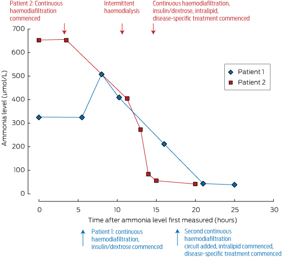
These measures should be initiated under the guidance of a metabolic physician, and in an intensive care unit where agitation or coma can be managed. Ammonia levels can be rapidly reduced by dialysis; its removal is dependent on flow rates, making intermittent haemodialysis the most effective method of clearance, as shown in Box 2. For this reason, we advocate intermittent dialysis rather than continuous venovenous haemodiafiltration for early ammonia control in the emergency setting.

While severe neurological impairment at the start of treatment is of great concern, this should not in itself be a reason to withhold treatment, as good neurological recovery is possible. This is illustrated by the case report of a middle-aged patient who recovered, despite decorticate posturing when therapy was initiated.5

We advocate early assessment of ammonia levels in patients with an unexplained altered conscious state, or when their cognitive disturbance seems disproportionate to any concurrent systemic illness. Many of the necessary elements of care can be initiated in non-tertiary intensive care units. Initiation of treatment at the hospital of presentation is essential, as this is a medical emergency; neurological outcome and survival are critically dependent on the timing of intervention. If recognised early and treated appropriately, the prognosis for neurological recovery is good.

Lessons for practice

  • Urea cycle disorders may first present in adulthood, unmasked by triggers such as systemic illness, increased protein load, surgery or corticosteroids.

  • Assessing ammonia levels is a simple but critical test in patients with unexplained impaired consciousness.

  • A session of intermittent haemodialysis is highly effective for rapid ammonia control, and superior to continuous haemodiafiltration for rapid correction.

  • Emergency treatment of hyperammonaemia should be undertaken early to prevent devastating neurological injury.

Box 1 –
Case histories of the two patients


Patient 1: 24 years old, male

Medical history

  • Obstructive sleep apnoea; no notable family history; high-functioning individual
  • Likely precipitant: intraoperative dexamethasone (8 mg) during nasal septoplasty

Progress

  • Vagueness and lethargy 48 h after operation, progressing over 24 h to incoherence
  • Intubated 12 h later for severe agitation
  • GCS declined to 5–6 over next 48 h; ammonia level, 334 μmol/L (RR, < 50 μmol/L); disease-specific treatment started
  • Raised intracranial pressures 6 h later (dilated pupils with cerebral oedema on CT brain), leading to decompressive craniectomy complicated by frontal haematoma, requiring evacuation
  • Prolonged intensive care unit and hospital stay

Outcome

  • Persistent minimally conscious state (at 22 months)
  • Discharged to nursing facility
  • Biochemical analysis of plasma and urine consistent with OTC deficiency (elevated urine orotic acid; plasma glutamine level high; plasma ornithine, citrulline and arginine levels low)
  • Genetic testing confirmed OTC gene mutation associated with OTC deficiency

Patient 2: 39 years old, male

Medical history

  • Chronic knee pain; no notable family history; high-functioning individual
  • Likely precipitant: cortisone injection into knee for knee pain

Progress

  • Headache, nausea, epigastric pain, blurred vision and incoordination 48 h after injection
  • Progressed over next 48 h to confusion, slowed speech
  • Progressive decline in GCS, requiring intubation
  • Seizure activity
  • Repeat CT brain showed cerebral oedema
  • Ammonia level: 652 μmol/L (RR, < 50 μmol/L); disease-specific treatment started; intracranial pressure monitor inserted; lack of control of intracranial hypertension; decision to palliate

Outcome

  • Proceeded to brain death and organ donation (except for liver donation: contraindicated)
  • Biochemical findings consistent with OTC deficiency (profound elevation of urine orotic acid, plasma glutamine level high, arginine level low)
  • Genetic testing confirmed OTC gene mutation associated with OTC deficiency

GCS = Glasgow coma score; RR = reference range; CT = computed tomography; OTC = ornithine transcarbamylase.

Box 2 –
Ammonia levels in our two patients, and their response to treatment*


* Note the rapid rate of decline of serum ammonia levels in Patient 2 achieved after initiating intermittent haemodialysis (about 10 hours after first measurement).

Time to shut down the acute care conveyor belt?

A rapid response system may be an appropriate model for meeting the urgent need for more suitable care for patients at the end of life

Hospitals can be dangerous places where people can unexpectedly die. Hospitals can also be dangerous places because people are not allowed to die. When they eventually die, it can be a prolonged and demeaning experience.1,2

The population of the world is increasing. People are living longer. An increasing number of aged people are spending their last few days, weeks or months in acute hospitals,3 many of whom will die in intensive care units (ICUs).4 Almost a third of Americans will spend time in an ICU during the last month of their life.4 However, most people want to die at home, not in an acute care hospital.

Discussions around the end of life (EOL) are ubiquitous and the term can be interpreted in many different ways. For the purposes of this article, we limit the term EOL to older people with significant comorbidities who, based on existing evidence, have less than 1 year to live. If EOL care in acute hospitals is one of the largest contributors to health care costs and if our society does not want it, how did it happen and how can we manage EOL care more appropriately and in line with what people want?

The acute care conveyor belt

The current situation is akin to older people with illness being placed on a conveyor belt, beginning in the community and eventually taking them to an acute hospital and then possibly to an ICU. Dying and EOL care have slowly and almost imperceptibly become medicalised. As we have developed impressive ways of keeping people alive, it has become very difficult to exercise the choice to die naturally and not be surrounded by machines and well-meaning people.

When people suffer sudden injury or illness in the community, an ambulance is often called, whether the illness is potentially reversible or simply a minor deterioration in someone with only days to live. The conveyor belt operates largely because of uncertainty of the patient’s prognosis, the lack of practical and readily available alternatives, and the failure of people to have stated their wishes regarding EOL care. Even if people’s wishes are expressed clearly, uncertainty can be a barrier to those wishes being carried out. Patients with severe chronic diseases marked by acute exacerbations become used to the idea that the medical system can always rescue them from life-threatening deteriorations. Of course, rescue is sometimes possible, until it is not. Well-meaning clinicians may convince patients that there is a large, potentially reversible component to their deterioration and that they deserve every chance to leave hospital alive. As a result, the rate of emergency admissions to hospitals and ICUs is dramatically increasing.57 Many of these patients are in the last year of life.6 Once patients are in the emergency department, it is easier to admit them to hospital rather than to embark on the time-consuming task of discussing options with them and their caregivers and finding a more appropriate place of care.6

After admission to the hospital, patients are confronted with further pressures to keep them on the conveyor belt. Hospital medicine has become increasingly specialised and fragmented.8 Hospital specialists are experts in their own particular part of the patient’s clinical status. However, the population of patients in acute hospitals has changed and elderly patients rarely have single-organ problems.9 They are now older, with multiple comorbidities. The ability to diagnose dying patients or to identify patients at the EOL, or understand the bigger picture of the patient’s circumstances, is often overlooked by single-organ specialists.10 All in all, the management of the dying process is not done well in acute hospitals.1,2

The final step in this process is to an ICU, where drugs and machines can, in theory, prolong life, no matter what the ultimate prognosis is.

The conveyor belt is also maintained and oiled by those who make the decision about whether the value of further medical intervention is futile. The diagnosis of dying, as defined by the well worn tenet “medicine at this time has nothing more to offer”, has moved from being made by general practitioners in community settings to hospital-based specialists. Deaths in ICUs are rarely sudden and unexpected.11 They are usually orchestrated as a result of withdrawing and withholding treatment after it has been decided, together with the patient and his or her carers, that further medical intervention has nothing else to give. Often, the underlying “disease” or combination of medicalised problems is simply the end result of normal and expected ageing.

Even in the ICU, the decision to continue active management may not be appropriate, as many of the survivors of the ICU do not leave hospital alive and many survivors of intensive care die within 12 months of discharge.12 For the cohort that does survive and leave hospital, there is a high incidence of a severe decrease in quality of life and of symptoms similar to those of posttraumatic stress disorder.13,14 Even if patients at the EOL survive the hospital intervention, little may have been done to improve posthospital survival or quality of life. Apart from the failure of the hospital system to recognise when people are at the EOL, there are other drivers pushing them along the conveyor belt. Society is bombarded with daily reports of medical miracles.15 There is little honest discussion about ageing and dying. Physicians seem to be complicit in this. It is sobering to reflect on the finding that most patients with terminal cancer may not be aware that the palliative chemotherapy they receive is unlikely to be curative.16

Rapid response systems and end-of-life care

Rapid response systems (RRSs) were established as a patient safety system to improve patients’ outcomes in acute hospitals.17 Their key features are vital sign and observational abnormalities which identify seriously ill patients and, in turn, trigger an urgent response by an individual or team with the appropriate skills, knowledge and experience to deal with any hospital emergency.17

Initially, they were established to identify seriously ill patients with potentially preventable illnesses. However, an RRS will also identify patients who are predictably and normally dying.18 Up to a third of all RRS calls are for patients who require limitations of treatment as a result of being at the EOL.18 This has important implications. It tells us that acute hospitals do not necessarily recognise patients at the EOL.9 The rapid response team becomes the surrogate “dying” team. The poor prognosis of the patient then has to be brought to the attention of the admitting team. It does not seem acceptable that patients only become aware of their parlous state when they are so close to death. The inappropriate management of patients at the EOL not only largely contributes to the unsustainable cost of health care but it is also not in the interest of patients and their carers to be denied rational and personal choices based on the prognosis.

Another approach to patients at the end of life

Managing patients in acute hospitals who are at the EOL may benefit from an approach similar to that used in RRSs. This would require identification of the patient followed by an appropriate response. There are currently attempts to identify such patients.19 Whatever tool is developed must deal with uncertainty, which is an integral part of medicine. However, a prognostic tool would equip the patient and the caring physicians with information such as the possibility of dying within, say, months or a year. We would not consider withholding poor prognostic information from a 20-year-old patient with a terminal brain tumour. The estimate of exactly how long that patient had to live would be uncertain in terms of weeks, months or even years in some instances. However, the same honest discussions usually do not occur with aged, frail patients who have had multiple admissions to hospital and who may have a similar prognosis to that young patient with the brain tumour.

It is early days in predicting prognosis in frail older people, but there are features (such as weight loss, significant decrease in mobility, gait speed, increasing weakness and exhaustion) which may indicate that a person has a high probability of having less than 12 months to live.19 Once a patient is identified as being in an at-risk group, there would need to be an urgent and appropriate response. As with RRSs, the response to the seriously ill, deteriorating patient would need to involve a clinician with appropriate skills, knowledge and experience, as well as the time to carry out lengthy and complex discussions with the patient and his or her carers. Currently, most admitting teams do not have that level of training.18 Moreover, the home team is often busy caring for the more conventional aspects of medical care. One could argue that we need an extensive rethink of our undergraduate and postgraduate training in order to give every clinician, including attending doctors and nurses, the skills to appropriately manage EOL care. Alternatively, or until we undergo that radical retraining, a response could be in the form of someone specifically trained in EOL care such as a palliative care nurse.

The response would obviously begin with involvement of the patient and their carers. Discussions would then be centred on the patients’ wishes, such as whether they want to be continually admitted to acute hospitals or whether they would prefer more home and community-based care. It would be essential to involve GPs in continuing management plans. These choices and the patient’s condition may, of course, change with time.

Other responses could include the offer of palliative care, perhaps in parallel with continued active treatment. Choices would include how patients would like to spend their last few months. Where they would like to spend them? What sort of support do they have? And what support do they need?

During these discussions, there would also need to be close communication with others in the palliative care team as well as members of the admitting team.

Conclusion

The current medicalisation of dying can be compared with the medicalisation of birthing in the 1950s, when mothers’ legs were put up in stirrups, babies were delivered and removed from their mothers and put in a large room together with other babies, and mothers were only allowed to see their babies for feeding. And, of course, fathers were excluded from the process altogether. Perhaps there is hope that the baby boomers, who have changed the way society operates in many ways, will demand greater choice and involvement in the way that their dying is handled.

We seem to have lost the ability to be honest with society about the limitations of modern medicine and to recognise that people will inevitably age and die badly. Currently, many patients at the EOL are placed on a conveyor belt where futile treatment and less than full disclosure have replaced genuine and appropriate care for patients and their carers.

New treatment to overcome peanut allergies in children

By Jane Trembath, Southern Health News / 4th of November, 2015

This story was first published by The Lead, South Australia.

A new study is successfully helping children to overcome peanut allergies by exposing them to peanuts and desensitising them to their allergy.

For the past four years, paediatric allergist Dr Billy Tao has been developing a novel two-step desensitisation process at Flinders Medical Centre (FMC) in South Australia.

The first step involves boiling peanuts for an extended length of time to make them less allergenic.

The boiled peanuts are given to patients to partially desensitise them, and then once the patient shows no signs of allergic reaction, roasted peanuts are given to the children to increase their tolerance in the second step of the process.

Dr Tao said the low-cost and effective two-step process resulted in less adverse events than previously used single-step desensitisation methods – also known as oral immunotherapy.

“With traditional methods, a lot of people ingesting increasing amounts of roasted peanut flour or similar products start to react – so much so that many have to drop out and can’t finish the treatment,” Dr Tao said.

The FMC trial is carried out over a year or longer and includes patients aged between 10 and 15 years.

Of the 14 participants, 10 have already completed the first step and are now eating varying amounts of roasted peanuts, while four continue to eat boiled peanuts and are progressing well.

“One patient who had to be administered three adrenaline injections after consuming peanuts is now eating several roasted peanuts every day without problems,” Dr Tao said.

Studies show the number of children living with peanut allergy appears to have tripled between 1997 and 2008, and as many as one in every 200 children will have severe allergy to nuts.

Allergy symptoms can vary from very mild (including tingling mouth, puffy lips and welts around the mouth) to moderate symptoms (facial swelling, body rash, runny nose and red eyes, abdominal pains and vomiting); while severe reactions include trouble breathing, looking pale and unwell, and anaphylaxis.  Very occasionally death may result from a most severe reaction. 

Dr Tao’s idea for hypo-allergenic (less allergenic) nuts to be consumed first was based on an observation by German researcher Professor Kirsten Beyer, who in 2001 noted that peanut allergies were less prevalent in China than the western world because the Chinese ate boiled peanuts rather than peanut butter or roasted peanuts. She found that boiling peanuts for 20 minutes made them less allergenic than roasted peanuts.

Dr Tao said that a partnership with Dr Tim Chataway, Head of the Flinders Proteomics Facility, and Professor Kevin Forsyth from the FMC Paediatrics Department, proved that peanuts boiled for at least two hours were less allergenic and the pair designed a study using this immunotherapy approach.

Dr Tao hopes his research could one day be carried out in a doctor’s clinic and then at home and avoid the need for hospital-based treatment.

However he strongly warned people against ‘do-it-yourself’ desensitisation at home and stressed that patients should be seen by an allergist and individual care plans developed.

Among those who have already undergone Dr Tao’s new desensitisation method is 16-year-old Shehan Nanayakkara, who was diagnosed with a severe peanut allergy at the age of three.

“We first realised Shehan had an allergy when friends gave him a peanut butter sandwich and he had to be rushed to hospital…there have been many accidents since then,” father Asanka said.

“During one round of allergy testing he ended up in the Intensive Care Unit – that time I thought I’d lost him.

“I approached Dr Tao to help and at first Shehan ate boiled peanuts, working his way up to consuming 13 a day, and now he eats five normal roasted peanuts daily, mixed in with his meals.

“It’s been a big relief because children and teenagers don’t care too much about what they eat and just eat whatever, and there has always been that worry that something might happen – now we can relax a bit because Shehan has some tolerance.”


[Correspondence] Fighting the ethnic fire with collaboration: a paediatrician’s perspective

April 28, 2015, was a beautiful day on the shore of Jaffa, Israel, where the Peres Center for Peace is located. We, a group of Israeli and Palestinian doctors, met there for a workshop focused on stabilisation of acutely ill or injured children. The workshop was led by Bruce Lister of Brisbane, Australia, and a strong, dedicated group of international and local faculty members. After the opening words and greetings by Lars Andersen, the European Union’s ambassador to Israel, and a later visit by former Israeli President Shimon Peres, we started 2 days of intensive training.

Deadly attacks raise fears of breakdown in rules of war

Governments and armed groups are being pressured to ensure the safety of patients and health workers in conflict zones amid a spate of high-profile attacks that have left dozens dead and injured.

The World Medical Association, the International Committee of the Red Cross, the World Health Organisation and several other peak health groups have jointly called on national governments and non-state combatants to adhere to international laws regarding the neutrality of medical staff and health facilities, and ensuring this commitment is reflected in armed forces training and rules of engagement.

The call follows the shelling of a Red Cross/red Crescent hospital in Yemen on Sunday, and the release of the initial results of a Medecins Sans Frontieres investigation into the deadly bombing of its hospital in Kunduz which found that there was no fighting occurring in or around the facility at the time of the attack, and that all armed groups, including the Afghan Army and the US Defence Department, had been given the accurate GPS coordinates of the hospital.

MSF said when the hour-long attack began soon after 2am on 3 October, 140 of its staff were at the hospital – including on 80 on duty – and they were treating 105 patients.

The humanitarian charity said at least 30 people were killed in the bombing raid, including 10 patients, 13 staff and seven whose remains were burnt beyond recognition. One MSF staff member and two patients are still missing and presumed dead.

Describing the attack in chilling detail, the charity reported that the intensive care unit, which was full at the time, was the first part of the hospital to be bombed. Several patients burned alive in their beds, a doctor had his leg blown off and a nurse had his arm virtually severed. One MSF staffer was decapitated by shrapnel, and several people were shot from the air as they attempted to flee the burning building.

As soon as the attack start, MSF made multiple calls to the Afghan Army and US armed forces, both in Kabul and to the Defence Department in Washington DC.

MSF International President Dr Joanne Liu said the internal review confirmed that MSF rules, including its strict ‘no weapons’ policy was in force and respected at the time of the attack, that the charity was in full control of the facility, that there were no armed combatants within the hospital compound, and there was no fighting from or in its vicinity before the airstrikes.

“We were running a hospital treating patients, including wounded combatants from both sides – this was not a ‘Taliban base’,” Dr Liu said.

The MSF President said the incident showed the deadly consequences of any ambiguity about how international humanitarian law applied to medical work in war.

“What we demand is simple: a functioning hospital caring for patients, such as the one in Kunduz, cannot simply lose its protection and be attacked,” Dr Liu said. “The attack…destroyed out ability to treat patients at a time when we were needed the most.

“We need a clear commitment that the act of providing medical care will never make us a target. We need to know whether the rules of law still apply.”

The United States and Afghan governments are yet to announce whether they will consent to an International Humanitarian Fact Finding Commission inquiry into the bombing.

But the Kunduz attack has nonetheless added to the urgency for action to be taken to ensure the safety of medical staff and hospitals in combat zones.

The International Committee of the Red Cross (ICRC), through its Health Care in Danger project, recorded 2398 attacks on health workers, facilities and ambulances in just 11 countries between January 2012 and the end of last year.

Policy and Political Affairs Officer for the ICRC’s Australian mission, Natalya Wells, said such attacks were not new, and were virtually a daily occurrence.

Ms Wells often health workers were caught in the cross-fire, particularly as a result of indiscriminate attacks in urban areas.

But she said that on occasion they were also being deliberately targeted, underlining the need for all combatants to respect the Geneva Conventions.
Ms Wells said that through the Health Care in Danger project, the ICRC was working with governments, armed forces and non-state combatants to improve awareness of, and respect for, laws and conventions around the protection of patients, health workers and medical facilities, particularly in conflict zones.

As part of the effort, governments attending the 32nd International Conference of the Red Cross and Red Crescent between 8 and 10 December are expected to back a resolution reaffirming their commitment to international humanitarian law and a prohibition on attacks on the wounded and sick as well as health care workers, hospitals and ambulances.

In addition, Ms Wells said the ICRC had held meetings with 30 non-state combatant groups from four continents about international humanitarian law and the rules of armed conflict.

The discussions have included incorporating knowledge of these conventions into their training, backed by sanctions for any breaches.

Promisingly, Ms Wells said that so far “one or two” non-state armed groups, though not signatories to the Geneva Conventions, have discussed creating a similar code of conduct for their forces.

Adrian Rollins


Signs workforce planning getting back on track

It’s been a chequered time for medical workforce planning in recent years.

Health Workforce Australia (HWA) was a Commonwealth statutory authority established in 2009 to deliver a national and co-ordinated approach to health workforce planning, and had started to make substantial progress toward improving medical workforce planning and coordination. It had delivered two national medical workforce reports and formed the National Medical Training Advisory Network (NMTAN) to enable a nationally coordinated medical training system.

Regrettably, before it could realise its full potential, the Government axed HWA in the 2014-15 Budget, and its functions were moved to the Health Department. This was a short-sighted decision, and it is taking time to rebuild the workforce planning capacity that was lost.

NMTAN is now the Commonwealth’s main medical workforce training advisory body, and is focusing on planning and coordination.

It includes representatives from the main stakeholder groups in medical education, training and employment. Dr Danika Thiemt, Chair of the AMA Council of Doctors in Training, sits with me as the AMA representatives on the network.

Our most recent meeting was late last month, and the discussions there make us hopeful that NMTAN is finally in a position where it can significantly lift its output, contribution and value to medical workforce planning.

In its final report, Australia’s Future Health Workforce, HWA confirmed that Australia has enough medical school places.

Instead, it recommended the focus turn to improving the capacity and distribution of the medical workforce − and encouraging future medical graduates to train in the specialties and locations where they will be needed to meet future community demands for health care.

The AMA supports this approach, but it will require robust modelling.

NMTAN is currently updating HWA modelling on the psychiatry, anaesthetic and general practice workforces. We understand that the psychiatry workforce report will be released soon. This will be an important milestone given what has gone before.

Nonetheless, it will be important to lift the number of specialties modelled significantly now that we have the basic approach in place, so that we will have timely data on imbalances across the full spectrum of specialties.

The AMA Medical Workforce Committee recently considered what NMTAN’s modelling priorities should be for 2016.

Based on its first-hand knowledge of the specialities at risk of workforce shortage and oversupply, the committee identified the following specialty areas as priorities: emergency medicine; intensive care medicine; general medicine; obstetrics and gynaecology; paediatrics; pathology and general surgery.

NMTAN is also developing some factsheets on supply and demand in each of the specialities – some of which now available from the Department of Health’s website (http://www.health.gov.au/internet/main/publishing.nsf/Content/nmtan_subc…). I encourage you to take a look.

These have the potential to give future medical graduates some of the career information they will need to choose a specialty with some assurance that there will be positions for them when they finish their training.

Australia needs to get its medical workforce planning back on track.

Let’s hope that NMTAN and the Department of Health are up to the task.

Pressure for independent inquiry of deadly US hospital attack builds

Pressure is mounting on the United States Government to agree to an independent inquiry into its attack on a hospital in Afghanistan that that left 22 people dead following the activation of a rarely-used international investigative body.

The International Humanitarian Fact-Finding Commission (IHFFC), established under the Geneva Conventions, has written to both the US and Afghanistan governments to offer its services for an independent inquiry following a complaint from medical charity Medicins Sans Frontieres, (MSF) which operated the hospital.

US President Barack Obama has issued a public apology for the bombing, and his Government has initiated its own inquiry. But Mr Obama has been steadfast in resisting calls for arms-length investigation, and is considered unlikely to accept the Commission’s offer.

Neither the US nor Afghanistan are member states of the Commission, which has no power to compel their participation.

“It is for the concerned Governments to decide whether they wish to rely on the IHFFC,” the Commission said. “The IHFFC can only act based on the consent of the concerned State or States”.

President Obama has assured that his Government would conduct a “transparent, thorough and objective” inquiry into the tragedy.

But MSF claims the attack could amount to a war crime and must be investigated independently.

“We have received apologies and condolences, but this is not enough. We are still in the dark about why a well-known hospital full of patients and medical staff was repeatedly bombarded for more than an hour,” said Dr Joanne Liu, MSF International President. “We need to understand what happened and why.”

Dr Liu said her organisation was determined to uncover how the attack had occurred, and to hold those responsible to account.

“If we let this go, as if it was a non-event, we are basically giving a blank cheque to any countries who are at war,” Dr Liu said. “If we don’t safeguard that medical space for us to do our activities, then it is impossible to work in other contexts like Syria, South Sudan, like Yemen.

Twenty-two people, including 12 MSF staff, were killed in the hour-long US airstrike, which was called in as Afghan Army units fought to regain control of the city from Taliban insurgents.

MSF nurse Lajos Zoltan Jecs survived the attack and detailed scenes of carnage at the hospital, which was filled with patients at the time.

“I cannot describe what was inside. There are no words for how terrible it was. In the Intensive Care Unit six patients were burning in their beds,” Mr Jecs said.

He told of how surviving medical staff – many badly shaken and traumatised by the blasts – worked frantically to save patients as well as their own colleagues.

“We did an urgent surgery for one of our doctors. Unfortunately he died there on the office table. We did our best, but it wasn’t enough,” he said. “We saw our colleagues dying. Our pharmacist – I was just talking to him last night and planning the stocks – and then he died there in our office.”

President Obama called Dr Liu to apologise for the attack after the US military admitted responsibility.

The attack occurred despite the fact that MSF had given all warring parties the GPS coordinates of the hospital.

Outrage over the attack was heightened when the US initially appeared to claim it was a necessary and legitimate use of force, before later characterising it as a mistake.

MSF said that “any statement implying that Afghan and US forces knowingly targeted a fully functioning hospital – with more than 180 staff and patients inside – razing it to the ground, would be tantamount to an admission of a war crime,” MSF Australia President Dr Stewart Condon and Executive Director Paul McPhun said. “There can be no justification for this abhorrent attack.”

“Medecins Sans Frontieres reiterates its demand for a full, transparent and independent international investigation to provide answers and accountability to those impacted by this tragic event.”

Adrian Rollins

[Clinical Picture] Cerebral fat embolism after bone fractures

After a forklift accident, a 44-year-old man was admitted to our hospital in July, 2014, with fractures of the right fibula and tibia, the left femur, and the pelvis. A proximal femoral nail antirotation was done to stabilise the left femur. 1 h after the operation, he developed acute respiratory failure, requiring reintubation and admission to the intensive-care unit. CT angiography of the thorax showed bilateral ground-glass opacities and distinct nodular opacities (figure), without pulmonary embolism or aspiration.

Deadly hospital attack could be war crime: MSF

Medical charity Medicins Sans Frontieres says the United States bombing of a hospital in strife-torn Afghanistan could be a war crime, and has insisted it be investigated by an independent commission despite assurances from President Barack Obama that his Government would conduct a “transparent, thorough and objective” inquiry into the tragedy.

As horrific accounts continued to emerge of the devastation wrought by the US bombing of the MSF-operated hospital in the Afghan city of Kunduz, MSF International President Dr Joanne Liu said her organisation was determined to uncover how the attack had occurred, and to hold those responsible to account.

“If we let this go, as if it was a non-event, we are basically giving a blank cheque to any countries who are at war,” Dr Liu said. “If we don’t safeguard that medical space for us to do our activities, then it is impossible to work in other contexts like Syria, South Sudan, like Yemen.

Twenty-two people, including 12 MSF staff, were killed in the hour-long US airstrike, which was called in as Afghan Army units fought to regain control of the city from Taliban insurgents.

MSF nurse Lajos Zoltan Jecs survived the attack and described scenes of carnage at the hospital, which was filled with patients at the time.

“I cannot describe what was inside. There are no words for how terrible it was. In the Intensive Care Unit six patients were burning in their beds,” Mr Jecs said.

He described how surviving medical staff worked frantically to save patients as well as their own colleagues.

“We did an urgent surgery for one of our doctors. Unfortunately he died there on the office table. We did our best, but it wasn’t enough,” he said. “We saw our colleagues dying. Our pharmacist – I was just talking to him last night and planning the stocks – and then he died there in our office.”

President Obama called Dr Liu to apologise for the attack after the US military admitted responsibility.

According to Fairfax Media, White House spokesman Josh Earnest said the US leader told Dr Liu that a US investigation would “provide a transparent, thorough and objective accounting of the facts and circumstances of the incident. And that, if necessary, the President would implement changes to make tragedies like this one less likely to occur in the future.”

But charity has insisted the attack be subject to an independent investigation, and has called for the International Humanitarian Fact-Finding Commission, which has been dormant since its creation under the Geneva Conventions in 1991, to be activated.

The attack occurred despite the fact that MSF had given all warring parties the GPS coordinates of the hospital.

Outrage over the attack was heightened when the US initially appeared to claim it was a necessary and legitimate use of force, before later characterising it as a mistake.

MSF has said the attack could amount to a war crime, and must be fully and independently investigated.

“Any statement implying that Afghan and US forces knowingly targeted a fully functioning hospital – with more than 180 staff and patients inside – razing it to the ground, would be tantamount to an admission of a war crime,” MSF Australia President Dr Stewart Condon and Executive Director Paul McPhun said. “There can be no justification for this abhorrent attack.”

“Medecins Sans Frontieres reiterates its demand for a full, transparent and independent international investigation to provide answers and accountability to those impacted by this tragic event.”

Adrian Rollins