×

Vocational training of general practitioners in rural locations is critical for the Australian rural medical workforce

The known In efforts to reduce the longstanding geographically inequitable distribution of Australian GPs, current policy requires that 50% of GP vocational training (registrar) positions are located in rural or remote areas. 

The new We identified a strong association between rural training pathways and subsequent rural practice, and it is intensified by a rural origin effect. Despite some attenuation over time, these associations remained strong up to 5 years after vocational registration. 

The implications Ongoing support for rural GP vocational training opportunities and the selection of rural origin medical students are critical components of GP workforce policy. 

The geographically inequitable distribution of the Australian medical workforce continues, and rural and remote general practitioner positions are largely filled by international medical graduates (IMGs).1 This dependency persists despite substantial government efforts to stimulate recruitment and retention of Australian-trained GPs in rural areas. Recent government initiatives have included a large increase in the number of federally supported medical school places for students, and supporting medical education and training in rural communities through the Rural Clinical Training and Support (RCTS) program.1,2 A quota for the proportion of domestic students with a rural background selected by medical schools (at least 25%) has also been introduced, and rural clinical exposure during undergraduate and pre-vocational medical training programs has increased. In addition, Australian policy now requires that 50% of GP vocational (registrar) training occurs outside metropolitan areas.1 This policy is based chiefly on research that has indicated that a positive educational experience in rural settings, targeted training of GP registrars for rural practice, and clear pathways to rural practice are the most effective incentives for interesting a GP in a rural career.3,4 Doctors accepted into GP training are selected into either the Rural Pathway or the General (mostly metropolitan) Pathway, with about 50% of candidates allocated to each.5

Evidence for the effectiveness of these interventions for increasing rural recruitment and retaining Australian medical graduates in rural areas has accumulated. Ranmuthugala and colleagues6 reported that evidence for the effectiveness of increased rural exposure during undergraduate medical training on the uptake of rural practice was inconclusive, but Wilkinson and colleagues7 found that postgraduate rural GP training had a stronger association with rural practice uptake than rural exposure during undergraduate training (although the availability of rural GP postgraduate training was low at the time of this study because the number of rural training positions was limited). More recent empirical data810 and data on intentions collected at training completion11,12 suggest moderate improvement in the uptake of rural practice by students who have participated in RCTS programs. However, as reported in three literature reviews on the recruitment and retention of medical practitioners in rural areas3,13,14 and as lamented in a recent letter to the Medical Journal of Australia,15 there remains a large evidence gap as to the effectiveness of rural exposure during vocational training programs. A review of the outcomes of the regionalised Australian General Practice Training Program16 found that only 27% of former Rural Pathway registrars remained in rural practice after 7 years. In addition, several North American studies have produced limited quantitative evidence of associations between vocational training in a rural primary care setting and subsequent rural practice.1720

The geographic origin of doctors also has an impact on their commencing rural practice, with convincing evidence about a strong link between an individual’s rural upbringing and their subsequent decisions about a rural career.21,22 The consistency of the reported association between GPs having a rural background and their choosing a rural career suggests that their origin is a critical factor in making this decision, regardless of vocational training location. Our study therefore aimed to investigate the association between vocational training location and the subsequent choice of practice location for newly registered GPs, including the effect of a rural background.

Methods

This study was based on data from the Medicine in Australia: Balancing Employment and Life (MABEL) study, conducted by the Centre for Research Excellence in Medical Workforce Dynamics (https://mabel.org.au/). MABEL is a national longitudinal survey that collects annual data from a panel of doctors, with a regular small participation top-up. The first wave of the MABEL study (2008) invited the entire medical workforce to participate, and 10 498 doctors (19.4% of the medical population) completed the initial survey, including 17.7% of GPs. There has subsequently been an annual 70–80% study retention rate. Further participants (generally recently graduated, non-specialist hospital doctors or IMGs newly registered in Australia) are added to the MABEL pool each year.

Our study analysed data from MABEL waves 1 to 7 (2008–2014), and was restricted to respondents who had completed their GP vocational training and were transitioning to independent practice. The transition year for a GP was identified from MABEL data on the basis of their participation in GP registrar training and details of newly completed medical qualifications. Data for IMG GPs — defined as those who had completed their initial medical training outside Australia and New Zealand — were analysed separately.

Rural origin and work location

Rural origin was defined for doctors trained in Australia or New Zealand as their having resided for at least 6 years in a rural area before the age of 18 years. Each doctor’s work location was geocoded in each MABEL wave to a specific town or suburb, then classified as metropolitan or rural. Rural location was defined as including Australian Standard Geographic Classification Remoteness Areas (ASGC-RA) 2 to 5;23 it was self-defined for New Zealand-trained doctors. Vocational training location was defined in two ways: as rural or metropolitan by work location in the year the doctor completed their training (final training location), and as an aggregate of work locations in the 2 to 3 years preceding their completion of training.

Statistical analysis

Four cohorts were defined by a combination of origin type and final training location: rural origin/rural training, metropolitan origin/rural training, rural origin/metropolitan training, and metropolitan origin/metropolitan training. For comparison purposes, IMGs were separately divided into two cohorts: rural training and metropolitan training.

A secondary (sensitivity) analysis defined four cohorts by multiple training locations: rural training only; completed training in a rural area, but also had some metropolitan training; completed training in a metropolitan area, but also had some rural training; metropolitan training only.

For each cohort, the proportions of GPs working in rural and metropolitan locations were calculated for each of the first 5 years after they had completed their vocational training. Rurally trained GPs were further classified according to whether they were working in the same or a different rural community from that in which they completed their vocational training; a buffer of 20 kilometres was allowed.

Separate generalised estimating equation (GEE) models with a logit link function and exchangeable correlation structure were used to test associations between vocational training pathways and subsequent work location for the four primary cohorts (non-IMGs only) for each of the 5 years after completing vocational training. Adjustments were made for four additional demographic variables during each particular year: sex, age, living with a partner, and having dependent children. A further variable — whether the GP was rurally bonded (contracted to work for part of their early career in rural locations) in a particular year — was included in each regression model. These models were repeated for the four secondary cohorts, with rural origin as an additional covariate; its multi-year cohort definitions limited analysis to 4 outcome years. All calculations were performed in StataSE 12 (StataCorp).

Ethics approval

The MABEL study was approved by the University of Melbourne Faculty of Business and Economics Human Ethics Advisory Group (reference, 0709559) and the Monash University Standing Committee on Ethics in Research Involving Humans (reference, CF07/1102 – 2007000291).

Results

During the 7-year study period, 610 doctors completed their GP vocational training and commenced in at least one subsequent work location. The demographic characteristics of these GPs are summarised in Box 1. Just under half of the local graduates (ie, those who graduated in Australia) trained in the Rural Pathway, and about one quarter were of rural origin (consistent with current policy requirements for GP training posts and medical student intakes); fewer than 10% were rurally bonded. Most local medical graduates were women, most lived with a partner, and almost 40% had dependent children. The proportions of IMGs who trained in the Rural Pathway, were men, were aged 35 years or more, lived with a partner, or had dependent children were higher than for local medical graduates (Box 1).

Box 2 summarises the practice location as an independent GP for the four primary cohorts of local medical graduates for each of the 5 years following their completion of vocational training. There were very strong and sustained associations between final vocational training location type and subsequent practice location for the rural origin/rural training and metropolitan origin/metropolitan training cohorts; 74–91% and 87–95% respectively remained in their origin/training type during their first 5 post-training years. Moreover, 61–70% of the rural origin/rural training cohort practised in the same rural community in which they trained during the first 4 years after completing their vocational training. Outcomes for GPs from cohorts 2 and 3 also showed a clear pattern: initially, these GPs generally remained in their final vocational training location type, but there was subsequently a gradual move in work location toward their origin type. The career patterns of rurally trained IMGs was similar to those of metropolitan origin/rural trained local graduate GPs, with a gradual move in work location toward metropolitan areas during the 5 years after vocational registration (Box 3).

The rural training pathway, regardless of childhood location, was highly significantly associated with subsequent rural practice. The odds of rural practice for each of the rural training cohorts of GPs decreased with time, but a strong and highly significant association was nevertheless retained across the 5 years. Unsurprisingly, rural bonding and rural origin were positively associated with rural practice. Higher age was also associated with rural practice, while there were no consistent statistically significant associations between practising in a rural location and sex, or with having a partner or dependent children (Box 4).

Secondary analysis, using the multiple year training location definition, confirmed the importance of rural training, particularly that of the final GP training year (Box 5).

Discussion

We have provided empirical evidence for the contribution of rural vocational training, in combination with the selection of rural origin students, to the Australian rural GP workforce. This is highly significant for rural workforce policy, as the Australian government requires that more than half of Australian GP vocational training positions be located in rural areas; our study allows an opportunity to assess the effect on the workforce of these policies.1

We found that training in the rural training pathway and the trainee having a rural background were each strongly associated with early career rural practice. The strength of the association between vocational training location and choosing rural practice remained strong and statistically significant up to 5 years after completing GP training for doctors of either rural or metropolitan origin (primary cohorts 1 and 2). Sustained rural practice was very strongly linked with the combination of a rural origin and rural training, but this cohort alone is unlikely to provide a sustainable rural GP workforce while only 25% of Australian-trained doctors are of rural origin, as about 30% of the Australian population live in rural or remote areas.

Most mixed rural/metropolitan origin/training GPs (cohorts 2 and 3) subsequently practised in a same location type as that in which they trained, although some gradually returned to their origin type. Diminution of the pathway effect over time is perhaps expected, as 50% of GP registrar training positions are in rural areas but about 75% of young doctors are of metropolitan origin. Other research has found that work location changes are most likely during early career stages,24 when personal circumstances, including relationships with spouses and dependents, are more fluid. The secondary analysis confirmed the strong influence of rural training on subsequent rural practice, especially location during the final year of vocational training. Together, these findings suggest that the periods leading up to and immediately following vocational training are critically important windows of opportunity for ensuring that appropriate policies optimise recruitment of GPs for rural practice and their subsequent retention.25,26

The largest cohort, metropolitan origin doctors undertaking GP training in metropolitan areas (cohort 4) largely remained in metropolitan practice. Further, there was no evidence that rural origin Australian doctors were more likely than metropolitan origin doctors to choose general practice as their specialty (unpublished MABEL data). Consequently, metropolitan origin doctors continue to remain the major source of non-IMG rural GPs, making cohort 2 (metropolitan origin/rural training) critical for the rural GP workforce. This cohort is nearly twice the size of cohort 1, and the association with rural practice was much stronger than for those in the metropolitan pathway (cohort 4). However, more than 50% of cohort 2 had moved to metropolitan practice after 5 years, further highlighting the importance of targeted retention initiatives focused on this cohort.

The odds of members of the smallest cohort (cohort 3: local medical graduates with a rural background who undertook their training in metropolitan areas) practising in rural areas was three times that for metropolitan origin/metropolitan training GPs, although the association was statistically significant only from 3 years after completing vocational training. However, the odds were much lower than for the rural origin/rural training cohort 1, highlighting the importance of the rural training pathway.

A key limitation of this study is that it cannot establish cause and effect. There is probably a strong self-selection bias, in that the rural training pathway attracts those who are interested in a rural career. Further limitations include the use of a self-selected cohort, the participants of the MABEL survey, who represent 15–18% of all Australian GPs. While the panel design of our study enabled individual tracking of doctors over a 7-year period and application of GEE (logit) modelling, the observed patterns, particularly in the mixed origin/training cohorts, suggest that these doctors have not yet decided on their long term preferred work location, and it is therefore difficult to accurately predict outcomes at, for example, 10 or 20 years. Additionally, vocational training location was primarily defined for the purposes of this study as the location of the trainee in the year they completed their training, as this was considered to be the most influential year for subsequent practice location. Our secondary analysis partially examined this aspect by separately analysing GPs who had undertaken vocational training in a mix of rural and metropolitan locations. Further, our key focus was on the joint effects of rural origin with rural/metropolitan training pathways. This necessitated a focus on GPs who had completed their medical degrees in Australia or New Zealand, despite IMGs comprising a considerable proportion of the rural GP workforce in Australia (more than 50% in some regions). Finally, this study used a binary measure of rurality (metropolitan v non-metropolitan) that may not adequately adjust for the substantial heterogeneity in the attractiveness to GPs of different rural and remote Australian locations. It is possible that more nuanced measures of rurality, including multiple levels of remoteness and population size, might have identified different associations for the four cohorts.27

Conclusion

Our study analysed the best available Australian longitudinal data about individual GPs to provide new quantitative evidence of a strongly positive association between rural GP vocational training location and subsequent rural practice, even after adjusting for the influence of rural origin. This evidence supports the objectives of existing policies that require at least 50% of GP training to occur in rural locations, and that at least 25% of medical students should be of rural origin. While Australia strives to reduce its reliance on IMG GPs for the rural workforce, this aim requires long term improvements in the rural recruitment and retention of Australian-trained GPs. Ongoing support for rural GP vocational training opportunities is a critical component of rural GP workforce policy in Australia.

Box 1 –
Demographic characteristics of participating doctors at the time they completed general practitioner vocational training

Local medical graduates

International medical graduates


Number

467

143

Rural Pathway (year of training completion)

221 (47.3%)

101 (70.6%)

Rural origin

118 (25.3%)

NA

Sex (women)

322 (69.0%)

74 (51.8%)

Age, median

32 years

41 years

Age, ≥ 35 years

153 (32.9%)

125 (89.9%)

Living with a partner

335 (72.7%)

119 (83.2%)

Has dependent children

179 (39.4%)

119 (83.8%)

Rurally bonded

35 (7.5%)

NA


NA = not applicable. Percentages exclude missing data for local medical graduates (age, 2; living with partner, 6; dependent children, 13) and international medical graduates (age, 4; dependent children, 1).

Box 2 –
Final vocational training location and general practice location for local medical graduates during the first 5 years after completing general practitioner vocational training

Time since completion of training

Location of practice

(1) Rural origin/rural training

(2) Metropolitan origin/rural training

(3) Rural origin/metropolitan training

(4) Metropolitan origin/metropolitan training


Number of GPs

78 (17%)

143 (31%)

42 (9%)

204 (44%)

1 year

Same rural

70%

54%

Other rural

20%

22%

18%

5%

Metropolitan

10%

25%

82%

95%

2 years

Same rural

62%

42%

Other rural

24%

31%

30%

13%

Metropolitan

14%

27%

70%

87%

3 years

Same rural

68%

24%

Other rural

15%

42%

35%*

11%

Metropolitan

18%

34%

65%*

89%

4 years

Same rural

61%

25%

Other rural

30%

29%

46%*

9%

Metropolitan

9%

45%

54%*

91%

5 years

Same rural

42%*

15%

Other rural

32%*

33%

33%*

9%

Metropolitan

26%*

52%

67%*

91%


* Groups with fewer than 20 participants.

Box 3 –
Final vocational training location and general practice location for international medical graduates during the first 5 years after completing general practitioner vocational training

Time since completion of training

Location of practice

Rural training

Metropolitan training


Number of GPs

101 (71%)

42 (29%)

1 year

Same rural

81%

Other rural

6%

4%

Metropolitan

13%

96%

2 years

Same rural

57%

Other rural

17%

8%

Metropolitan

26%

92%

3 years

Same rural

49%

Other rural

10%

0*

Metropolitan

41%

100%*

4 years

Same rural

45%

Other rural

21%

18%*

Metropolitan

34%

82%*

5 years

Same rural

53%*

Other rural

7%*

20%*

Metropolitan

40%*

80%*


* Groups with fewer than 20 participants.

Box 4 –
Odds of local medical graduates practising in a rural location during the first 5 years after completing general practitioner vocational training

Odds ratio (95% confidence interval)


1 year post-GP training

2 years post-GP training

3 years post-GP training

4 years post-GP training

5 years post-GP training


Primary cohorts

(1) Rural origin/rural training

159 (45–558)

65 (27–158)

48 (22–102)

50 (24–106)

52 (24–111)

(2) Metropolitan origin/rural training

68 (26–175)

32 (16–60)

28 (16–51)

23 (13–41)

24 (13–43)

(3) Rural origin/metropolitan training

2.8 (0.7–11)

2.4 (0.9–6.2)

2.9 (1.2–6.7)*

3.3 (1.5–7.4)

3.5 (1.5–7.9)

(4) Metropolitan origin/metropolitan training

1.00

1.00

1.00

1.00

1.00

Age (for each 1-year increase in age)

1.06 (1.00–1.13)*

1.04 (0.99–1.08)

1.04 (1.00–1.08)*

1.05 (1.01–1.08)*

1.04 (1.01–1.08)*

Sex (reference: men)

1.00 (0.48–2.1)

0.9 (0.5–1.6)

1.03 (0.6–1.7)

0.8 (0.5–1.4)

0.8 (0.5–1.4)

Living with a partner

0.8 (0.3–1.9)

0.9 (0.5–1.7)

0.9 (0.5–1.7)

0.98 (0.6–1.7)

0.9 (0.6–1.5)

Has dependent children

1.8 (0.8–4.1)

1.9 (1.06–3.3)*

1.4 (0.9–2.3)

1.3 (0.9–2.0)

1.3 (0.9–1.9)

Rurally bonded

5.1 (1.2–22)*

3.5 (1.1–11)*

3.8 (1.4–11)*

3.7 (1.4–10)*

3.6 (1.3–10)*


Odds ratios from generalised estimating equation (logit) model: * P < 0.05; † P < 0.01.

Box 5 –
Odds of practising in a rural location for each of the 4 years after completing general practitioner training for local medical graduates

Odds ratio (95% confidence interval)


1 year post-GP training

2 years post-GP training

3 years post-GP training

4 years post-GP training


Secondary cohorts

(1) Rural training only

92 (27–312)

49 (21–115)

41 (19–88)

29 (14–59)

(2) End training rural, with some metropolitan training

17 (5–58)

11.6 (4.6–29)

11.5 (4.9–26)

9.9 (4.3–23)

(3) End training metropolitan, with some rural training

0.94 (0.09–9.4)

2.8 (0.8–9.4)

2.9 (1.00–81)

2.7 (0.96–7.9)

(4) Metropolitan training only

1.00

1.00

1.00

1.00

Rural origin

4.1 (1.3–13)*

2.0 (0.9–4.3)

2.1 (1.02–4.1)*

2.5 (1.3–4.9)

Age (for each 1-year increase in age)

1.2 (1.04–1.3)

1.08 (1.01–1.16)*

1.07 (1.01–1.14)*

1.05 (1.00–1.12)

Sex (reference: men)

0.9 (0.3–2.4)

0.8 (0.4–1.7)

0.9 (0.5–1.9)

0.8 (0.4–1.5)

Living with a partner

0.6 (0.2–2.1)

1.1 (0.5–2.6)

1.1 (0.5–2.4)

1.07 (0.5–2.1)

Has dependent children

0.6 (0.2–2.0)

1.3 (0.6–2.7)

1.09 (0.6–2.1)

1.02 (0.6–1.8)

Rurally bonded

2.0 (0.4–10)

2.21 (0.6–7.8)

3.8 (1.2–13)*

3.6 (1.1–11)*


Odds ratios from generalised estimating equation (logit) model: * P < 0.05, † P < 0.01.

Composite reliability of workplace-based assessment for international medical graduates

The known Workplace-based assessment (WBA) of the performance of doctors has gained increasing attention. The reliability of individual assessment tools has been reported in previous studies. 

The new We analysed the composite reliability of a toolbox of WBA instruments in assessing international medical graduates (IMGs). For five case-based discussions, 12 Mini-Clinical Examination Exercises and six multisource feedback assessments, the composite reliability coefficient was 0.899 (standard error of measurement, 0.125). 

The implications The reliability of WBA for assessing the performance of IMGs is excellent. WBA can also be used for performance assessment in other settings. 

The purpose of this article is to report the value of workplace-based assessment (WBA) for evaluating international medical graduates (IMGs). Most countries have systems for assessing IMGs. Fundamental to these systems are robust assessment procedures that assess their fitness to practise, and they typically include written multiple choice question tests and objective structured clinical examinations.1,2 The virtue of standardised tools is that the assessment is similar for all candidates. Despite having been validated,3 however, they do not assess proficiency in actual practice. The disadvantage of standardised assessment is its questionable relevance to real world clinical practice; it has been suggested that the “standardisation of final, licensing, and fitness to practise examinations may make educationalists weep with joy, but there is no clear evidence that it makes for better doctors.”4 Could we perhaps do better?

In recent years, WBA has become more prominent in medical education. Its purpose is to assess proficiency in an authentic clinical environment, principally because what doctors do is more important than what they know, for both patients and society.57 Many postgraduate training bodies are implementing WBA strategies,8 and several undergraduate programs are already using some of its tools, particularly the Mini-Clinical Evaluation Exercise (mini-CEX), case-based discussions (CBDs), multisource feedback (MSF), and Directly Observed Procedural Skills (DOPS). The philosophy underpinning WBA is the assessment of several domains by multiple assessors over a period of time, with feedback built into each encounter.9 This form of assessment can track the progress of the trainee, for which reason WBA is described as “assessment for learning”, rather than the traditional “assessment of learning”.6 Although originally developed for formative assessment (for feedback and training), these tools have been used in programmatic assessment (in which multiple assessment tools are used to comprehensively assess a doctor or student in a well designed program) and can be used for summative purposes (to determine whether a candidate has passed or failed a course or program).

We hypothesise that WBA has the potential to provide more relevant assessment of IMGs. When applied to assessing their fitness to practise, WBA must be robust and validated for this purpose. Earlier studies of WBA for IMG assessment found that WBA is acceptable to the candidates, assessors and the health care system,10 and our earlier study found that it is also cost-effective.11 Although feedback from supervisors and staff indicate that WBA candidates are ready to work at a satisfactory level, there has been no reliability study of WBA for IMG assessment.

Moreover, studies of the reliability of WBA instruments typically focus on a single instrument, but, in practice, assessment information is pooled across methods. We therefore need a multivariate estimate of the composite reliability of the WBA toolbox, as first suggested by Miller and Archer6 and undertaken by Moonen-van Loon and colleagues in a recent study of domestic graduates.12 They found that combining the information from several methods meant that smaller samples were adequate (ie, fewer individual tests of each type).

The question therefore arises: what is the composite reliability of WBA when used for high stakes (ie, critical) assessment of IMGs? Our study estimated the composite reliability of a WBA program in Australia. Although trainees receive supervisor reports during most training programs, this has been found to “under-call under-performance”, as the reports are prepared by a supervisor who is also the assessor (both coach and referee).13 Since this was a routine assessment and many of the IMGs had completed different assessment forms, we only analysed the newer tools: mini-CEX, CBDs and MSF.4,8

Methods

All IMGs who wish to practise in Australia (except those who qualified in the United Kingdom, the United States, Canada, Ireland or New Zealand) must pass the Australian Medical Council (AMC) examination. This assessment consists of a multiple choice examination and an English proficiency assessment, followed by a clinical examination (16 objective structured clinical examination stations) in an examination centre.14

In 2010, we established a program to assess these doctors with WBA as an alternative to the AMC clinical examination. Many IMGs are accorded temporary registration that allows them to work in areas where there is a workforce shortage while waiting for the AMC clinical examination. This waiting period is often long. To be eligible for WBA in our program, the candidates had to pass the English and multiple choice question examinations, and be employed for the duration of the program (6 months). If the candidate passed our assessment, they were eligible for AMC certification. Our assessment program is accredited by the AMC.15

Data collection

Data were collected from June 2010 to April 2015. During this 5-year period, IMGs employed in Hunter New England Health, both in urban and rural areas, completed 970 CBDs, 1741 mini-CEX and 1020 MSF assessments, managed and administered by the Centre for Medical Professional Development Unit in Newcastle. There were 103 male and 39 female candidates from a broad range of countries (Afghanistan, Argentina, Bangladesh, Belgium, Burma, China, Egypt, Fiji, Germany, India, Indonesia, Iran, Iraq, Italy, Jordan, Kenya, Malta, Malaysia, Nepal, the Netherlands, Norway, Pakistan, Papua New Guinea, Romania, Sierra Leone, South Africa, Sudan and Ukraine).

In total, 99 assessors rated the CBD and mini-CEX assessments. The MSF assessors were nominated by the IMGs, and the assessment forms were sent, collected and analysed by the central office; the forms were de-identified when results were provided to the candidates. Over the 5-year study period, more than half the assessors attended follow-up re-calibration and feedback sessions. Ongoing review of the quality of the program was undertaken by an independent group consisting of clinical academics, educationalists and administrators who oversaw the governance of the program. All candidates attended similar calibration sessions of about 3 hours each. Several different assessors assessed each IMG during the 6-month period. All results were recorded on the assessment forms and sent directly to the central office. The data were stored at a secure site.

WBA instruments (Appendix)

The assessment consisted of 12 mini-CEX examinations, five CBD examinations and one set of MSF data, and each candidate was assessed by at least six assessors. The mini-CEX assessments in medicine, surgery, women’s health, paediatrics, emergency medicine and mental health were blue-printed (designed) to reflect the AMC examination. The assessment level was appropriate for the first postgraduate (intern) year.

The mini-CEX, originally developed in the US to guide learning, is used to assess clinical performance in authentic clinical situations.16 The IMG was assessed in six disciplines and various competencies, and scored on a scale of 1 to 9; 1–3 corresponds to unsatisfactory performance, 4–6 to satisfactory performance, and 7–9 to superior performance. Case complexity and global rating were marked during the constructive feedback. The CBDs, which assess the candidate’s record-keeping and clinical reasoning, were scored on a similar scale.17,18 To pass, the IMG had to achieve a satisfactory result in eight of 12 mini-CEX and four of five CBDs, and to pass the MSF (with the average score of 3).

For the MSF, the IMG nominated three medical and three non-medical colleagues (eg, nurse, social worker, pharmacist) with whom they had worked extensively during the assessment period to complete an assessment form. The IMG also completed a self-assessment form. An MSF assessment form consisted of 23 questions with statements on aspects such as professionalism, communication, and requesting help when in doubt, and were scored on a 1–5 scale.6,19

We used the overall score of the mini-CEX and CBD assessments and the average scores of all scored items in the MSF assessments. When including the MSF assessments in the WBA toolbox, the scores were linearly transformed to a 1–9 score by multiplying the average score by 2 and subtracting 1.

We did not include the self-assessment results from the candidates in the MSF data, as this item was for their own reflection and not for evaluation of performance by external assessors. Reports from supervisors were not included in the analysis, as they have been found to be unreliable.13

Data analysis

All mini-CEX, CBD and MSF assessments for a candidate over a period of 6 months were extracted. The secured records were analysed in SPSS 23 (IBM). For each assessment, we calculated the average score to analyse the reliability of the various WBA tools, as well as the composite reliability of the tools as a group.

Reliability analysis

Reliability analysis assesses the reproducibility or consistency of WBA scores, and therefore provides an indication on how well we can differentiate between the levels of performance (scores) of the IMGs. Generalisability theory takes into account different sources of variance and is therefore considered a useful framework for estimating the reliability of complex performance assessments.20 It generates a reliability coefficient with a range of 0 to 1. When providing a high stakes assessment based on a combination of several low stakes assessments, a reliability coefficient of 0.8 is generally regarded as acceptable.21

The numbers of assessments and assessors varied between IMGs, and each assessor assessed a different set of IMGs. The facet (ie, source of variation) of average assessment scores (i) is therefore nested within the facet of IMGs (p), leading to the generalisability design i:p. For each WBA tool, we estimated variance components using analysis of variance with type I sums of squares (ANOVA SS1). The absolute error variance for the decision study on the separate WBA instruments is calculated by dividing the estimate of the variance component σ2 (i:p) by the harmonic mean for each instrument. The harmonic mean was preferred to the arithmetic mean because the number of assessment scores differed between IMGs, and because the harmonic mean tends to reduce the effect of large outliers (ie, a single IMG with many assessments).22

Distinct from the separate univariate reliability of each WBA instrument, the composite reliability of all instruments as a toolbox is calculated using a D-study in multivariate generalisability theory.22 Each assessment score (i) is a score for exactly one assessment instrument, and the corresponding multivariate model is therefore i:p ; ie, the facet of IMGs (p) is crossed with the fixed multivariate variables (assessment instruments) and nested within the independent facet of assessment scores (i). The composite universe score and absolute error variances are determined by a weighted sum of the universe scores and absolute error variances of the individual assessment instruments. The weights can be optimised by multivariable optimisation to obtain an optimal composite reliability coefficient.12

Ethics approval

Ethics approval to collect and analyse the data was obtained from the Hunter New England Health Human Research Ethics Committee in 2010 (reference, AU201607-03 AU). All IMG candidates and assessors provided consent to use their de-identified data.

Results

Box 1 summarises the number of assessments and the number of IMGs tested during the study period, with mean scores (on a 1–9 scale), standard deviations, and harmonic means for each of the assessment types (average number of assessments).

Reliability of the individual WBA instruments

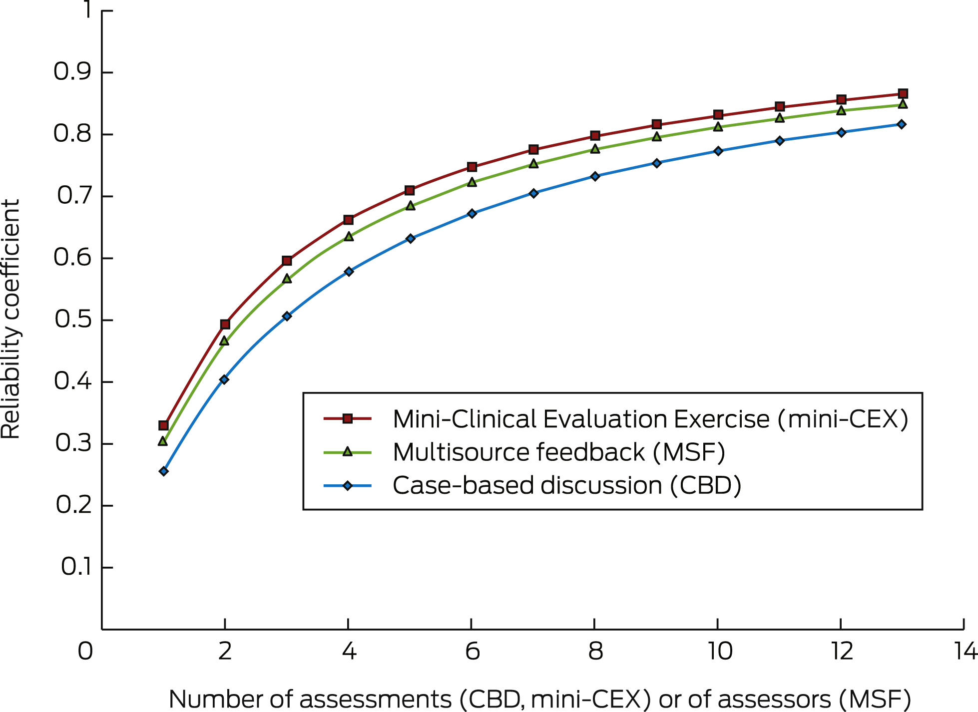
Box 2 presents the reliability coefficients according to the number of assessments (CBD and mini-CEX) or assessors (one occasion of MSF). The data were derived from the regular variance components for the true and error variance associated with individual assessment tools. The minimum number of assessments needed for a reliability coefficient of 0.8 was 12 for CBDs, nine for mini-CEXs and ten for MSFs.

Composite reliability of the WBA toolbox

As 5-point scale was used for the MSF assessments, but 9-point scales for the CBD and mini-CEX assessments, we performed two composite reliability studies: one that excluded the MSF assessments, and one that included them after linearly transforming their scores to a 9-point scale.

The reliability threshold of 0.8 could be attained by a combination of five CBD and five mini-CEX assessments, but also with three CBD and six mini-CEX assessments (Box 3). As described in the introduction, IMGs generally undergo 12 mini-CEX and five CBD assessments during the 6 months; this combination had a reliability coefficient of 0.886 and a standard error of measurement (SEM) of 0.144. The SEM estimates how average scores per assessment of an IMG were distributed around their “true” score (ie, performance level). However, the dataset indicated that typically more assessments of all types were undertaken than required, leading to a reliability coefficient of 0.895 and an SEM of 0.138 when using harmonic means of test numbers and optimised weights (Box 1).

Adding the six MSF assessments to the five CBD and 12 mini-CEX assessments slightly increased the reliability coefficient from 0.886 to 0.890, with an SEM of 0.131. Using harmonic means (Box 1) and optimised weights, we obtained a reliability coefficient of 0.899 and an SEM of 0.125 (Box 4).

Discussion

An assessment instrument for evaluating performance in a high stakes setting should have a reliability coefficient of at least 0.8. Our study found that our WBA program meets this criterion. The composite reliability we found is as good as or even better than that of most standardised assessments.23 Our previous studies have found the WBA program has good acceptability, educational impact, and validity.10 Taken together, our program therefore satisfies the criteria for a “good assessment” program.9

Further, when the components were used as part of a WBA toolbox, we achieved good reliability with fewer individual assessments.12 This may lead to changes in the procedure, reducing the workload for IMGs and assessors. It should be noted that all instruments in the toolbox meet the standards set by the AMC. They focus on different aspects of performance, but have similar assessment scales, and are applied by assessors adhering to the same assessment standard after calibration. These characteristics allow for the combination of the WBAs in one toolbox, allowing composite reliability scores to be calculated. It is interesting that when we searched for optimal weights for individual instruments in the aggregation for the composite score, the mini-CEX received the most weight, perhaps because the mini-CEX has the highest individual reliability (Box 2).

Assessment fatigue is a major problem in clinical assessment, and any program should aim to optimise the use of the assessors’ time.24,25 With fewer assessments, more people are likely to implement such a program. The current program was also highly acceptable to the IMGs because of the educational value inherent in the immediate constructive feedback.26

We examined the performance of IMGs in Australia, but the 5-year study period and the large number of assessments included in the dataset render it sufficiently rigorous that the results can probably be extrapolated to other programs. However, the level of calibration of assessors and the structure of the assessment instruments should be similar if comparable results are to be obtained.

Evaluating the performance of doctors (what they do) is more important than assessing their competency (what they know), as their performance during training and practice is more relevant to patients and society. This is especially important in the case of doctors educated in different medical training systems. WBA programs with multiple tools provide a reliable method for assessing IMGs and can be delivered in a well organised, blue-printed program that assures the breadth and depth of the assessment. Similar programs could have a huge impact on the performance of IMGs, potentially improving patient outcomes. However, we do not know whether the long term performance of candidates who undergo WBA is different from IMGs who pass the traditional examination, and comparison of these outcomes for the two pathways would be desirable.

Most postgraduate training programs are adopting WBA components. The tools used by the assessors have individual reliabilities greater than 0.8, and our study may contribute to designing an improved portfolio of assessment, with different assessment tools for achieving more rigorous performance assessment.

Box 1 –
Numbers of assessments and of international medical graduates tested during the study period, June 2010 – April 2015, and summary of the test scores

CBD

mini-CEX

MSF


Number of assessments

970

1741

1020

Number of international medical graduates

142

141

141

Mean number of assessments per graduate

6.8

12.3

7.2

Harmonic mean number of assessments

6.7

12.2

6.7

Mean test score

6.0

5.8

7.1

Standard deviation

0.7

0.6

0.7


CBD = case-based discussion; mini-CEX = Mini-Clinical Evaluation Exercise; MSF = multisource feedback.

Box 2 –
The reliability of the individual workplace-based assessment instruments

Box 3 –
Composite reliability when combining different numbers of Mini-Clinical Evaluation Exercises and case-based discussion assessments, with optimised weights

Shaded cells: reliability coefficient ≥ 0.8 (threshold for acceptability).

Box 4 –
Result of the D-study with equal and optimised weights for the different workplace-based assessment tools, using the harmonic means of numbers of assessments

CBD/mini-CEX


CBD/mini-CEX/MSF


Equal weights

Optimised weights

Equal weights

Optimised weights


Weights

0.500, 0.500

0.270, 0.730

0.333, 0.333, 0.333

0.240, 0.638, 0.122

Universe score

0.169

0.163

0.123

0.140

Error score*

0.025

0.019

0.018

0.016

Reliability coefficient

0.869

0.895

0.870

0.899

SEM

0.160

0.138

0.136

0.125


CBD = case-based discussion. mini-CEX = Mini-Clinical Evaluation Exercise. MSF = multisource feedback. * Calculated by dividing the covariance by the harmonic mean, summed for all instruments, divided by the number of different instruments.

The potential of workplace-based assessment of international medical graduates

The concept is attractive — but capacity may limit its practicality

For many years, Australia has relied on supplementing its medical workforce with doctors who have qualified outside Australia. Each year, about 2500 of these medical practitioners, known as international medical graduates (IMGs), seek general registration with the Medical Board of Australia. For many IMGs, this has included sitting the clinical examinations conducted by the Australian Medical Council (AMC) as part of the Standard Pathway for IMGs. The eligibility standard for registration is set at the expected level of an Australian medical graduate at the time they complete their internship.1 Concerns have been expressed about the accessibility of these examinations and the ability of IMG candidates to pass them. Some of these problems were highlighted during an inquiry in 2011–2012 by the House of Representatives’ Standing Committee on Health and Ageing, Lost in the labyrinth.2

Workplace-based assessments (WBAs) have been developed as alternatives to the Objective Structured Clinical Examination (OSCE) and other approaches for assessing clinical competence.3 They have the perceived advantage of allowing a single set of high stakes (summative) assessments in an examination environment to be replaced by multiple low stakes (formative) assessments conducted by supervising clinicians over a period of time and in the workplace. These methods have been progressively adopted in recent years by medical specialist colleges. The AMC commissioned a trial of WBAs as an alternative to their clinical examination in 2010, and have subsequently incorporated WBAs into the Standard Pathway.

An article by Nair and colleagues in this issue of the MJA evaluates the reliability of WBAs in this context.4 The usefulness of an assessment method relies on a number of psychometric criteria being fulfilled. These include the concepts of validity (does the assessment method reflect performance in practice?), reliability (is it reproducible and consistent?), feasibility (is it an efficient use of resources?), acceptability, and educational impact.5 WBAs are generally recognised as having high validity, as they are conducted in the workplace where the doctor is practising and are modelled on and executed as part of normal clinical practice.

Reports have previously been published about the feasibility and acceptability of WBAs for assessing IMGs, and on the reliability of a single method, the mini-clinical evaluation exercise (mini-CEX).68 A combination of assessment methods, however, allows different aspects of clinical practice to be assessed.3 The article by Nair and colleagues examined the reliability of such a combination (“composite reliability”), and found that it was good (reliability coefficient greater than 0.8).

Why is this important? In an examination process, reliability can be ensured by standardised processes, similar formats, and controlling the examiners, candidates and the examination environment. However, this often compromises the validity of the examination. The increased validity associated with WBAs, on the other hand, can affect reliability because of variations and pressures in the clinical environment, the fact that examinees are dealing with real patients, differences in assessment formats, and the lesser control over the candidates and assessors. A high degree of reliability for a combination of assessments indicates that candidates are being assessed in a standard and reproducible manner and to an equivalent standard of competence, comparable with the standard AMC examination process. The results reported by Nair and colleagues are, however, for a single program; for these findings to be generalised to other WBA programs for IMGs, further research may be required.

What do these results mean for assessing IMGs in the future? In 2015, 84 candidates participated in the WBA program, and 76 completed it successfully. In comparison, changes to the AMC processes and the establishment of the Vernon C. Marshall National Test Centre in Melbourne have allowed 2000 candidates to attempt the clinical examination over the same period (with about 590 completing it successfully).9 While WBAs have been shown to be feasible, affordable and reliable, they require resourcing and a commitment from the host institution. Further, candidates need to be recruited to a training position in a hospital offering the program. Investing in the program has rewarded some hospitals with improved recruitment and retention of practitioners, which is important in regional areas where this can be difficult. However, competition for training positions has increased; there are more than 3000 domestic medical graduates each year who need pre-vocational training.10

The AMC clinical examination offers access to a greater number of IMGs (possibly reflected in the lower rate of successful completions than for WBA). Australia continues to be an attractive destination for medical graduates from other countries, and the demand for assessment will therefore continue. WBAs will be a useful part of that assessment, but there are limits to the number of candidates who can be accommodated by this approach, especially when compared with the AMC examination.

The jugular veins: gateway to the heart

Inspection of the jugular veins provides a simple means of determining whether pressures in the right side of the heart are normal or elevated. With practice, clinicians can derive accurate and reliable information relevant to diagnosis and patient care.

Identifying a venous pulsation

It is not necessary to position the patient at precisely 45 degrees.1,2 If your patient is in a chair, examine them in that position. If they are on a couch or bed, examine them in the position that you find them.

Explain to the patient why you want to look at their neck. Traditionally, the right side of the neck has been used for jugular vein examination. However, it is often more difficult to see pulsations on the same side as you are positioned and, importantly, it is known that measurements made from the left side of the neck have similar accuracy.3 Further, inspection of the external, rather than internal jugular vein are also of similar accuracy.4 If you do use the ipsilateral side of the neck, try side-lighting with a torch or looking tangentially across the skin, rather than directly at it. Whichever side you use, and whichever vein, there must be visible pulsation at the top of the venous column. If there is no visible pulsation, do not use that vein as a manometer.

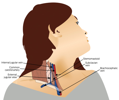
Ask the patient to turn their head slightly away from the side you are observing and focus on the area where the internal jugular vein is located — the anterior triangle (Box 1).

If you cannot see any pulsation at all, try lying the patient flatter or sitting them up — this may make a venous pulsation visible. If you still cannot see any pulsation, try sustained firm pressure in the upper abdomen. This is known as abdominojugular reflux and may transiently elevate a venous pulsation from below the clavicle and make it visible. If you have to do this to make a venous pulsation visible, it usually means that the right atrial pressure is not elevated.

When you identify a pulsation, decide whether it is arterial or venous. Box 2 shows the key distinguishing features.

If you decide that the pulsation is arterial, try abdominojugular reflux or changing the position of the patient to see if any additional pulsation appears.

If the veins of the neck seem distended but are non-pulsatile, sit the patient up at 90 degrees. This may make the pulsatile top of a venous column visible.

On most occasions, unless the patient is very obese, this systematic approach will allow confident identification of a venous pulsation. You can then use the pulsation to estimate right atrial pressure, whatever position the patient is in.

Estimating the right atrial pressure from observation of a jugular vein pulsation

When you identify a jugular vein pulsation, do not try and make a measurement in centimetres, just decide whether the pressure is normal or elevated. The simplest way to do this is as follows. If the top of the pulsating venous column can be seen to be more than 3 cm above the angle of Louis (sternal angle) in whichever way you have positioned the patient, this is highly predictive of an elevated right atrial pressure.1 Remember that clinical evaluation of the jugular vein pressure, just like ultrasound evaluation, typically underestimates the right atrial pressure.1 If you are confident that the jugular vein pressure is elevated, this reinforces the likelihood that the right atrial pressure is high.

What do I need to know about the waveform?

The jugular vein waveform is complex with three peaks — atrial contraction (a), ventricular contraction (c) and venous filling of the atrium (v) — and two troughs — atrial relaxation (x) and ventricular filling (y). Most clinicians can recognise the multiphasic quality of the venous pulsation but cannot confidently identify the specific peaks and troughs or their abnormalities. In real life clinical practice, this is of little importance. However, one abnormality of waveform is not uncommon. The video at www.mja.com.au demonstrates the giant v wave, which makes the venous pulsation almost look uniphasic and can mimic arterial pulsation if the steps described in Box 2 are not followed. This waveform is highly predictive of the presence of tricuspid regurgitation.5

Clinical value of jugular vein pressure estimation

In situations when accurate and multiple measurements of right atrial pressure are required — for example, the acutely unwell patient in a high dependency or intensive care setting — direct measurement by invasive (catheter) or non-invasive (ultrasound) means is usually preferred.

However, for the large numbers of patients cared for in ambulatory or general ward settings — particularly when heart failure is questioned as a diagnosis, or is known to be present and decisions about treatment are required — evaluation of the venous pressure by the method described here remains valuable. In the longer term, bedside ultrasound may supersede this technique. However, in the immediate future and in the absence of widespread access to such technology, bedside assessment of jugular venous pulsation is accurate and convenient, and continues to be a gateway to good clinical care of patients with heart disease.

Box 1 –
Jugular vein anatomy — the anterior triangle

Box 2 –
Features that help distinguish an arterial from a venous pulsation in the neck

Arterial pulsation

Venous pulsation


Appearance

Uniphasic, single

Multiphasic, undulating, flickering*

Effect of changing the position of the patient

None

May change its position in the neck

Effect of respiration

None

Falls on inspiration, rises with expiration

Palpation over the pulsation

Palpable

Impalpable (but beware of pressing too deeply, as you may feel the carotid)

Effect of gentle pressure at base of neck

None

Ceases

Effect of sustained pressure on the upper abdomen (abdominojugular reflux)

None

Transient rise


* The video at www.mja.com.au shows an exception to this general rule.

Streamlining ethics review for multisite quality and safety initiatives: national bariatric surgery registry experience

The current ethics review process is inappropriate for clinical quality registries

Rigorous methods for assessing and improving the quality of health care have proven difficult to develop by traditional research approaches.1 Clinical quality registries (CQRs) systematically collect an agreed minimum dataset of data across multiple sites on clinically relevant outcome measures. Data are analysed, comparing procedures, providers and institutions.2 Feedback to practitioners has been shown to drive performance improvement, especially if the data are perceived to be high quality.3

Because CQRs collect and store health information, protocols require human research ethics committee (HREC) approval to ensure that they comply with the Australian Privacy Act 1988 (Cwlth). Principle 6 of this Act states that stored personal information must not be used or disclosed for a secondary purpose unless patient consent is obtained or there is a permitted health situation. Section 16B of the Act defines the permitted health situations, which include research relevant to public health or public safety. The use or disclosure of personal information must be conducted under guidelines approved under section 95A of the Act. Current National Health and Medical Research Council guidelines state that ethical review is required at each contributing site for CQRs except where multi-institutional approval is in operation.4

Bariatric surgery is burgeoning in Australia. In 2016, it is estimated that there will be over 15 000 such procedures performed in Australia at a direct cost of over $225 million. Yet there are no evidence-based guidelines directing who should be offered this surgery, nor are there any long-term community data documenting its safety and efficacy in Australia.

The Obesity Surgery Society of Australia and New Zealand (OSSANZ) partnered with Monash University Department of Epidemiology and Preventive Medicine (DEPM) to establish a national registry of all bariatric procedures with the aim of filling these knowledge gaps. The pilot commenced in 2012 and national rollout commenced in May 2014 (with federal government funding).

The Bariatric Surgery Registry (BSR) collects information on each procedure performed, the devices used, changes in patients’ weight and diabetes status, and adverse events. Data are collected primarily from surgeons and are validated against hospital International Statistical Classification of Disease and Related Health Problems, 10th Revision, Australian Modification (ICD-10-AM) discharge codes. Because the BSR is tracking and storing identifiable sensitive health information longitudinally as well as cross referencing data points to external data sources, HREC review is required at every site contributing to the BSR.4,5

We believe that there are 164 hospitals undertaking bariatric surgery in Australia. As of 30 April 2015, there were 52 hospitals (31.7%) for which HREC approval for participation in the BSR had been obtained. Private hospitals accounted for 67% of these sites, 29% were public hospitals and an additional approval was received from both the Royal Australasian College of Surgeons and Monash University. Applications for high risk projects were requested from 31.4% of private hospital and 33.3% of public hospital HRECs. Seven sites (13.5%) provided approval through an affiliated site. Fifty sites (96%) had additional governance requirements and, in 76% of cases, this was a separate process. The median time from the first application to final approval was 86 days (range, 17–414 days). The maximum numbers of queries from or changes requested by an HREC was 67.

The process of obtaining ethical approval at these initial hospital sites cost the BSR $180 698.58 in salaries and $3474.97 per application. In addition, the BSR has had to pay five sites a total of $3927.00 for ethics approval application fees.

The number of CQRs in Australia is growing rapidly in response to community demands for better monitoring of health care outcomes.6 HREC review of registry processes is one way of ensuring that the rights of individuals participating in registries are protected and complying with the Privacy Act. However, as highlighted by our experience in rolling out the BSR, the lack of a consistent process for obtaining HREC approval across multiple sites for these quality and safety initiatives creates cost and slows implementation.

HRECs are typically set up to review research projects rather than quality and safety initiatives. Unlike clinical trials, which are hypothesis driven and in which patients are given the option of participating, CQRs attempt to ensure quality through benchmarking, and so need to recruit all patients who undergo a given intervention to avoid the risk of bias. Many HRECs are not familiar with these basic and essential differences, and this can lead to confusion and delays, as the processes for clinical trials and CQRs invariably differ.7 Based on the cost and time involved in obtaining even 31.7% of the required approvals for the BSR, we call for a bespoke national process for HREC review of CQRs. This would streamline implementation and reduce costs while still protecting patient’s privacy. Examples of such a process could be having all sites apply to a single national ethics committee, as in New Zealand, or implementing specific federal legislation protecting the transfer of information to and from approved CQRs.

News briefs

Ken Harvey awarded ANZAAS Medal

Dr Ken Harvey, renowned anti-pseudoscience activist and critic of regulatory agencies, has been awarded the Australian and New Zealand Association for the Advancement of Science (ANZAAS) Medal for 2016. Dr Harvey, who is adjunct associate professor in Monash University’s School of Public Health and Preventive Medicine, was presented with medal by Dr Malcolm Jenkins, the chair of ANZAAS, on 17 August. The medal is awarded each year for “services for the advancement of science or administration and organisation of scientific activities, or the teaching of science throughout Australia and New Zealand and in contributions to science that lie beyond normal professional activities”. Sir Gus Nossal and Sir Mark Oliphant are previous winners. Dr Harvey is an executive member of Friends of Science in Medicine (FSM), and has a national reputation as a strong champion of evidence-based medicine and treatment. “My interest in unethical promotion started in the 1970s when I was trying to contain hospital acquired antibiotic-resistant microorganisms,” Dr Harvey said. “I advocated the use of older, narrow-spectrum, more cost-effective antibiotics. The response of many pharmaceutical companies was, ‘You can’t afford to be wrong, use our latest, broadest-spectrum and most expensive antibiotics.’ A number of purveyors of complementary, alternative and integrative medicine also make unethical claims. So what to do? Marshal the evidence; flood the regulators with complaints, engage the media and agitate for policy change.” Congratulating Dr Harvey on his award, Professor John Dwyer, president of FSM said: “Ken Harvey is a champion for better public health in Australia. His efforts over many years have been focused on reducing the harm to consumers associated with misleading and even fraudulent promotion and use of treatments and medicines for which there is no scientific support.”

Zika virus tentacles reach further

As of 10 August, 69 countries and territories have reported evidence of mosquito-borne Zika virus transmission – 66 of them since 2015 – reports the World Health Organization. The latest to join the list include the United States, the Cayman Islands, and the Netherlands. Since February 2016, 11 countries have reported evidence of person-to-person transmission of Zika virus, probably via a sexual route. As of 10 August, 15 countries or territories have reported microcephaly and other central nervous system malformations potentially associated with Zika virus infection or suggestive of congenital infection. Canada is the latest country to report a case of congenital malformation associated with a travel-related case of Zika virus infection. The US Centers for Disease Control and Prevention has reported 15 live-born infants with birth defects and six pregnancy losses with birth defects with laboratory evidence of Zika virus infection. Since 10 August, 16 countries and territories have reported an increased incidence of Guillain-Barré syndrome (GBS) and/or laboratory confirmation of a Zika virus infection among GBS cases. Grenada is the latest country to report a case of GBS associated with a confirmed Zika virus infection.

Feel better by planting trees and taking up yoga

Planting trees or picking up litter is good for the environment and makes you feel better, but don’t cancel the health insurance just yet — the evidence for genuine health benefits is limited. That’s the finding of a review of 19 studies of outdoor environmental enhancement and conservation activities. People taking part in these activities perceived a range of benefits, including increased social contact and sense of achievement. It’s one more reason to support Clean Up Australia Day (doi: 10.1002/14651858.CD010351.pub2).

Similarly, asthma sufferers who want more control over their symptoms should consider yoga in addition to their usual medication. A recent review of 15 trials involving over 1000 men and women with mild to moderate asthma found that yoga exercise reduced the impact of asthma on quality of life and led to small improvements in symptoms. The effects on lung function and medication use were inconclusive (doi: 10.1002/14651858.CD010346.pub2).

Another recent asthma review sought to compare different durations and doses of oral steroids for relieving symptoms in people experiencing asthma attacks. Despite 18 trials involving over 2400 adults and children, only low quality evidence was found. Few studies could be combined in any meaningful way, making it difficult to tell whether longer or shorter courses or higher or lower doses are better or safer (doi: 10.1002/14651858.CD011801.pub2).

There’s no disguising the target demographic for football broadcasts with the constant reminder that even if men are not bald yet, they soon will be. But what of the less talked about female pattern hair loss? Twenty-five new studies have been added to the pile in the recent update of treatment options that now includes 47 trials of nearly 5300 women. There are many comparisons to navigate but, in short, minoxidil (either 2% or 5%) is more effective than placebo, finasteride is not, and laser comb therapy, although not rated as better than sham therapy by participants, did result in an important increase in hair growth (doi: 10.1002/14651858.CD007628.pub4).

For more on these and other reviews, visit the Cochrane Library at www.cochranelibrary.com.

Disparities in acute in-hospital cardiovascular care for Aboriginal and non-Aboriginal South Australians

The known Disparities in the treatment of Aboriginal and non-Aboriginal patients hospitalised with acute coronary syndromes have been reported. 

The new After adjusting for age and other factors, Aboriginal status was independently associated with lower coronary angiography rates. Angiography was more likely if family members or Aboriginal liaison officers were present. Revascularisation rates and prescription of medical therapies were similar for Aboriginal and non-Aboriginal patients who had undergone angiography. 

The implications The reasons for lower angiography rates among Aboriginal patients are complex, but equality of treatment can be achieved. Improving the hospital experience for Aboriginal patients is needed to reduce disparities in treatment. 

Coronary heart disease (CHD) contributes significantly to the 10-year life expectancy gap between Aboriginal and non-Aboriginal Australians.13 CHD mortality is estimated to be twice as high among Aboriginal Australians,3 accounting for 14% of all deaths of Aboriginal people.4 A higher incidence of acute coronary syndromes (ACS), particularly of acute myocardial infarctions (AMI), among Aboriginal Australians is a major contributor to premature mortality in this population. In 2006, the Australian Institute of Health and Welfare identified major disparities in the management of Aboriginal and Torres Strait Islander patients hospitalised with ACS between 2002 and 2003, including a 40% lower rate of coronary angiography, a 40% lower rate of percutaneous coronary intervention (PCI), and a 20% lower rate of coronary artery bypass graft surgery (CABG).5

Given the extent of the inequalities related to cardiovascular disease, there is clearly a need to identify and overcome problems that contribute to these health disparities. To expand knowledge in this area, our study examined the in-hospital management of Aboriginal people with an ACS, focusing on explaining these disparities. Our aims were: (i) to assess differences in the rates of angiography and subsequent revascularisation for Aboriginal and non-Aboriginal South Australians presenting with an ACS, taking into account age and other factors; and (ii) to explore the reasons for any disparities by undertaking a detailed review of individual hospital admissions.

Methods

This was an observational study of Aboriginal and non-Aboriginal patients presenting with an ACS to any public tertiary hospital in South Australia between January 2007 and December 2012. We undertook a retrospective analysis of hospital administrative data and a chart review of all admissions of Aboriginal people with an ACS during this period (matched with a control cohort of non-Aboriginal patients).

Establishment of the expert working group

Following the release of Better hospital care for Aboriginal and Torres Strait Islander people experiencing heart attack,6 it was recognised that contemporary SA-specific data were required for evaluating disparities in care in this state. Consequently, the Heart Foundation in SA, in collaboration with the SA Health Cardiology Clinical Network, established a research team to provide the data. The project was overseen by an expert working group with representation from the research team and each SA Health local health service, as well as Aboriginal health professionals.

Administrative data

To investigate differences in diagnostic angiography and revascularisation (PCI/CABG) rates in Aboriginal and non-Aboriginal people, hospital admissions for ACS to all South Australian public tertiary facilities, with separations between 1 January 2007 and 31 December 2012, were obtained from the Integrated South Australian Activity Collection (ISAAC). ISAAC records are coded using International Statistical Classification of Diseases and Related Health Problems, 10th revision, Australian modification (ICD-10-AM) codes. All records were based on separations (episodes of care) rather than individual patients. Records were included in the analysis if the principal diagnosis was AMI (ICD-10-AM, I21.x) or unstable angina (I20.x) and the admission was recorded as an acute episode of care with casualty/emergency department as the source of referral. To avoid contamination of the dataset, records for interhospital transfers were excluded. Records in which Indigenous status was not stated or was inadequately described were also excluded.

Chart review

To further examine the in-hospital management of Aboriginal patients with an ACS, a chart review was undertaken, using 1:1 matching and a standardised case report form. The hospital medical records for each separation recorded in the administrative data of patients who were identified as Aboriginal or Torres Strait Islander were extracted. They were matched as closely as possible with the next admission to the same hospital of a non-Aboriginal patient of the same age (within 10 years) and sex, and with the same principal diagnosis. Documentation of the medical decision-making process about whether the patient received invasive management was also reviewed; influences on this process were categorised as clinical or non-clinical (ie, patient-related factors).7 Data quality for the chart review was ensured by using trained abstractors and record re-abstraction for 10% of cases. Training included a detailed data dictionary and coding instructions, a testing phase with training examples, and individual education sessions on site before and during data collection.

Statistical analysis

Administrative data

Age at admission (mean, standard deviation) for Aboriginal and non-Aboriginal patients was compared with the Student t test. Comparisons of categorical endpoints for Aboriginal and non-Aboriginal patients are presented as percentages, and were assessed by logistic regression. Unadjusted and age-adjusted comparisons are reported as odds ratios with 95% confidence intervals (CIs). The primary outcomes included receiving diagnostic coronary angiography and, for patients for whom angiography was performed, PCI and CABG rates. To explore the association between invasive management and Aboriginal status, logistic regression models were fitted for each outcome. Modelling commenced with fully saturated models that included Aboriginal status and all remaining predictors with P < 0.20 as factors; non-significant variables (P ≥ 0.05) were then systematically removed from the models.

Chart review

Clinical outcomes for Aboriginal and non-Aboriginal patients are presented as percentages and compared by logistic regression as described above. Symptom duration in minutes (median, interquartile range [IQR]) was compared with the Wilcoxon rank-sum test. The primary outcome in the chart review was the prevalence of clinical and non-clinical factors associated with non-invasive management; outcomes for the two groups were compared by logistic regression, with unadjusted and age-adjusted results reported.

All tests were two-tailed (α = 0.05). All analyses were performed in Stata/IC 11.2 for Mac.

Ethics approval

This study was approved by the SA Health Human Research Ethics Committee (reference, HREC-13-SAH-89; 461/07/2014) and the Aboriginal Health Research Ethics Committee (reference, 04-13-516).

Results

Administrative data

During January 2007 – December 2012, a total of 13 843 separations with a principal diagnosis of ACS were recorded. Aboriginal status was not stated or inadequately described in 722 records (5.6%), leaving 13 071 admissions records for analysis, of which 274 (2.1%) referred to Aboriginal patients. The mean age of the Aboriginal patients was about 16 years lower than for non-Aboriginal patients, and a higher proportion were women (Box 1). The age distribution by Aboriginal status and sex is summarised in Appendix 1. Most patients presented with an AMI; the figure was similar for the two groups (57%), despite the younger age profile of the Aboriginal patients. Evaluation of additional diagnosis codes found a higher prevalence among Aboriginal patients of several cardiovascular risk factors and comorbidities, including diabetes, smoking, dyslipidaemia and renal failure (Box 1).

Analysis of hospital procedure codes indicated that 6069 separations (46.4%) included a diagnostic coronary angiogram during the admission for an ACS (Box 2). The proportion was similar for Aboriginal and non-Aboriginal patients, but age-adjusted analyses identified that Aboriginal patients were significantly less likely to undergo the procedure (Box 2). Box 3 shows the prevalence of coronary angiography by age group, indicating a significant disparity for Aboriginal patients in the 45–54 years (P < 0.001) and 55–64 years (P = 0.001) age groups.

For the entire cohort, patients who underwent coronary angiography were significantly younger than those who did not (mean [SD], 64.0 years [12.9] v 73.8 years [13.8]; P < 0.001), and a greater proportion were men (72% v 58%; odds ratio [OR], 1.87; 95% CI, 1.74–2.01; P < 0.001). Multivariable logistic regression identified eight independent factors significantly associated with angiography: Aboriginal status (OR, 0.4; 95% CI, 0.3–0.5; P < 0.001); age (as a continuous variable: OR [for each one-year increase in age], 0.9; 95% CI, 0.9–1.0; P < 0.001); AMI as principal diagnosis (OR, 5.8; 95% CI, 5.3–6.3; P < 0.001); sex (OR [men v women], 1.3; 95% CI, 1.2–1.4; P < 0.001); major cities (OR [v outer regional/remote/very remote area residence], 2.9; 95% CI, 2.3–3.7; P < 0.001); renal failure (OR, 0.5; 95% CI, 0.4–0.6; P < 0.001); heart failure (OR, 0.5; 95% CI, 0.5–0.6); P < 0.01); and airway disease (OR, 0.6; 95% CI, 0.5–0.7; P < 0.001).

After adjustment for age, the odds of Aboriginal patients undergoing PCI were significantly lower than for non-Aboriginal patients (Box 2). The between-groups odds ratios for undergoing CABG were significant in neither the unadjusted nor the age-adjusted analyses. However, when the analysis of interventional procedures was restricted to patients who underwent coronary angiography, PCI rates for Aboriginal and non-Aboriginal patients were similar, as were those of CABG and revascularisation overall (Box 2). For admissions that included a coronary angiogram, multivariable models assessing independent predictors of revascularisation (PCI, CABG) did not find an association with Aboriginal status.

Chart review

We reviewed medical record abstracts for 274 Aboriginal and 274 non-Aboriginal patients. Aboriginal patients were matched for sex with non-Aboriginal patients (57% were men), but were significantly younger (mean age [SD], 53.1 years [10.5] v 59.0 [10.4] years; P < 0.001). The number who arrived at hospital by ambulance was similar for Aboriginal and non-Aboriginal patients (148 [61%] v 149 [60%]; age-adjusted OR, 1.3; 95% CI, 0.9–1.8; P = 0.24), as was symptom duration (median time before hospital presentation [IQR], 160 min [80–415 min] v 165 min [80–419 min]; P = 0.62). The angiography rate among Aboriginal patients was lower than for non-Aboriginal patients in both the unadjusted and age-adjusted analyses (141 [51%] v 169 [62%]; age-adjusted OR, 0.5; 95% CI, 0.4–0.7; P < 0.001). Stratification according to the National Heart Foundation/Cardiac Society of Australia and New Zealand ACS guidelines8 indicated a similar risk burden for the patient groups (Appendix 2). The proportion of Aboriginal patients who underwent angiography was lower in each risk group, but the difference was statistically significant only for patients with high risk non-ST-elevation acute coronary syndrome (NSTEACS; 49% [59 patients] v 60% [64 patients]; age-adjusted OR, 0.5; 95% CI, 0.3–0.9; P = 0.02) (Box 4). Aboriginal patients who received care facilitated by an Aboriginal liaison officer were significantly more likely to have an angiogram (28% [27 patients] v 9% [10 patients]; OR, 3.9; 95% CI, 1.8–8.6; P = 0.001), as were Aboriginal patients who arrived at the hospital with an escort (43% [32 patients] v 24% [25 patients]; OR, 2.4; 95% CI, 1.3–4.6; P = 0.01).

The documentation on the decision not to proceed with angiography for Aboriginal and non-Aboriginal patients is summarised in Box 5. Non-clinical factors were more frequently cited to explain managing an ACS without angiography in documentation for Aboriginal patients than for non-Aboriginal patients. For more than one third of Aboriginal patients, the reason for choosing conservative management was unclear, compared with 10% for non-Aboriginal patients (Box 5).

At discharge, the prescription of guideline-recommended therapies for AMI patients was similar for Aboriginal and non-Aboriginal patients (Box 6). Regardless of Aboriginal status, secondary prevention therapies, including aspirin, β-blockers and cardiac rehabilitation referral, were prescribed significantly less frequently for those who did not undergo coronary angiography than for patients who did (Appendix 3).

Discussion

In this evaluation of patients attending the major tertiary facilities in SA for the treatment of ACS, the mean age of Aboriginal patients was about 15 years lower than for non-Aboriginal patients. Despite their being younger, high risk features were worryingly common, including higher rates of background cardiovascular risk factors and comorbidities. Importantly, we identified that the major difference in the in-hospital treatment of Aboriginal patients with an ACS was the rate of coronary angiography: after correcting for the effects of age, sex, principal diagnosis, comorbidities and remoteness, Aboriginal patients with an ACS were significantly less likely to undergo this diagnostic procedure. However, we also found that revascularisation (PCI or CABG) rates following angiography were similar for Aboriginal and non-Aboriginal patients. Similar to findings in the Northern Territory,9 Aboriginal and non-Aboriginal patients who had undergone angiography were prescribed evidence-based therapies on discharge at comparable rates. These findings show that equivalent treatment for Aboriginal and non-Aboriginal patients is achievable.

The lower age-adjusted odds of Aboriginal patients undergoing diagnostic angiography is concerning, but consistent with previous reports of lower rates of cardiac intervention in national5 and state-based datasets.10 Similar rates of CABG, but not of PCI, among Aboriginal and non-Aboriginal patients have been described.10 We, however, found similar rates of PCI for the two population groups; the reasons for this difference are unclear, but may include advances in PCI technology11 that have expanded the range of Aboriginal patients who can be treated with this intervention. Unlike other studies, we also undertook a restricted analysis of revascularisation rates that included only admissions where a diagnostic angiogram had been performed, thereby differentiating different sources of disparity; this analysis indicated that the major difference involved the decision about diagnostic angiography. Research in the United States exploring racial disparities in treating CHD also found that the greatest disparity was the referral to angiography.12,13

To further examine the causes of the disparity in angiography rates, we undertook a medical record review of all Aboriginal ACS admissions in the hospital data, matched with data for a non-Aboriginal cohort. This information provided insight into the medical decision-making processes for Aboriginal ACS patients and confirmed that a negative hospital experience, bias based on complex comorbidities or presumed adherence to medications that favoured conservative management, and patient choice were implicated in the difference in angiography rates.6,14,15

In 56% of cases in which Aboriginal patients did not undergo angiography, the decision was attributed to patient-related factors or no clear justification was provided, compared with 17% for non-Aboriginal patients. The rate of discharge against medical advice was high among Aboriginal patients who did not receive angiography (10.5%). This raises concerns about barriers to quality care for Aboriginal people, including poor engagement and communication, a lack of coordinated care, and inadequate cultural competence of health care providers.16,17 All of these factors can result in isolation, fear and disengagement by the patient.9

The importance of shared discussions and cultural support structures is highlighted by our findings, as Aboriginal patients who arrived at the hospital with an escort (family member or friend) were more likely to undergo angiography. Encouraging shared decision making and enabling systems that support a companion during the hospital stay are crucial for improving Aboriginal health care.14 Our data also strongly support the involvement of Aboriginal liaison officers, as their presence was associated with an increased likelihood of angiography. Although we cannot presume a direct causative relationship, these interactions are understood to improve communication and coordination, and to assist in alleviating fear.17

It has previously been reported that Aboriginal people with symptoms suggestive of a heart attack may delay presenting to a hospital.9 However, our findings suggest this situation may be improving, as symptom duration at hospital presentation was similar for Aboriginal and non-Aboriginal patients. Strategies that target early response and action may now be having an impact, with a number of culturally appropriate health resources and education tools available to patients and health care providers.

The major limitations to our study were that the data were drawn from one Australian state, and that the assessed records were based on hospital separations rather than individual patients. Further, patients receiving care in non-tertiary facilities were not included in our analysis. An evaluation of these patients, with a particular focus on comparing patients who were subsequently transferred with those who remained in a non-catheterisation facility, would be desirable. Our investigation could thus be strengthened by broader population coverage and by using linked data.

Our study provides important insights into the in-hospital treatment of Aboriginal patients with an ACS. We found a significant health disparity in the rates of coronary angiography for Aboriginal and non-Aboriginal patients that is not explained by the frequency of complex comorbidities in these populations. Hospitalisation for acute cardiovascular care can be distressing, and for Aboriginal patients this problem is compounded by limited understanding by health care workers of factors influencing Aboriginal health, leading to miscommunication that may reinforce negative perceptions, patient disengagement, and fear. In our study, engagement with the patient’s family and care facilitated by Aboriginal liaison officers each had positive impacts. While there are other contributing factors to disparities in treatment, health care workers and systems that facilitate a constructive hospital experience will improve the ability to provide effective care for Aboriginal patients.

Box 1 –
Clinical characteristics of Aboriginal and non-Aboriginal patients presenting with an acute coronary syndrome to South Australian tertiary hospitals, 2007–2012

Aboriginal patients

Non-Aboriginal patients

Odds ratio for Aboriginal patients


Unadjusted (95% CI)

P

Age-adjusted (95% CI)

P


Total number of separations

274

12 797

Age at admission (years), mean (SD)

53.1 (10.5)

69.6 (14.1)

< 0.001

Sex (women)

117 (43%)

4460 (35%)

1.4 (1.1–1.8)

0.01

2.7 (2.1–3.5)

< 0.001

Location*

Major cities

203 (74%)

10 730 (84%)

0.6 (0.4–0.7)

< 0.001

0.8 (0.6–1.1)

0.19

Inner regional

12 (4.4%)

1615 (13%)

0.3 (0.2–0.6)

< 0.001

0.2 (0.1–0.4)

< 0.001

Outer regional

25 (9.2%)

310 (2.4%)

4.1 (2.6–6.2)

< 0.001

2.6 (1.7–4.0)

< 0.001

Remote/very remote

33 (12.1%)

128 (1.0%)

14 (9.1–20)

< 0.001

9.2 (5.9–14)

< 0.001

Principal diagnosis

Unstable angina

115 (42%)

5403 (42%)

0.9 (0.7–1.2)

0.93

0.9 (0.8–1.3)

0.51

Acute myocardial infarction (NSTEMI or STEMI)

155 (57%)

7241 (57%)

1.0 (0.8–1.3)

0.99

1.0 (0.8–1.3)

0.71

Unspecified acute myocardial infarction

4 (1%)

153 (1%)

1.2 (0.5–3.3)

0.69

2.3 (0.8–6.5)

0.11

Cardiovascular risk factors

Hypertension

162 (59%)

7745 (61%)

0.9 (0.7–1.2)

0.64

1.3 (1.0–1.7)

0.20

Diabetes

121 (44%)

2737 (21%)

2.9 (2.3–3.7)

< 0.001

3.0 (2.4–3.9)

< 0.001

Current smoker

129 (47%)

2610 (20%)

3.5 (2.7–4.4)

< 0.001

1.2 (0.9–1.6)

0.18

Dyslipidaemia

86 (31%)

3074 (24%)

1.4 (1.1–1.9)

0.01

1.1 (0.8–1.4)

0.56

Family history of ischaemic heart disease

15 (5.5%)

477 (3.7%)

1.5 (0.9–2.5)

0.14

0.6 (0.4–1.1)

0.09

Cardiovascular comorbidities

Cerebrovascular disease

2 (1%)

179 (1.4%)

0.5 (0.1–2.1)

0.36

1.2 (0.3–5.0)

0.79

Peripheral vascular disease

3 (1%)

311 (2.4%)

0.4 (0.1–1.4)

0.17

0.5 (0.2–1.6)

0.27

Cardiomyopathy

7 (3%)

169 (1.3%)

2.0 (0.9–4.2)

0.09

2.0 (0.9–4.3)

0.09

Valvular heart disease

11 (4.0)%

791 (6.2%)

0.6 (0.3–1.2)

0.14

1.4 (0.7–2.6)

0.30

Heart failure

23 (8.4%)

1766 (14%)

0.6 (0.4–0.9)

0.01

1.7 (1.1–2.7)

0.02

Cardiogenic shock

3 (1%)

240 (1.9%)

0.6 (0.2–1.8)

0.35

1.0 (0.3–3.1)

0.96

Non-cardiovascular comorbidities

Renal failure

41 (15%)

1546 (12%)

1.3 (0.9–1.8)

0.15

3.6 (2.5–5.1)

< 0.001

Malignancy

3 (1%)

214 (1.7%)

0.7 (0.2–2.0)

0.46

1.1 (0.4–3.6)

0.83

Airway disease/asthma

6 (2%)

534 (4.2%)

0.5 (0.2–1.2)

0.11

0.8 (0.4–1.9)

0.64

Liver disease

2 (1%)

59 (0.5%)

1.6 (0.4–6.5)

0.52

1.7 (0.4–7.2)

0.47


NSTEMI = non-ST-elevation myocardial infarction; STEMI = ST-elevation myocardial infarction. * Classified according to Australian Bureau of Statistics, Postcode 2012 to Remoteness Area 2011 concordance (released 31 Jan 2013: http://www.abs.gov.au/AUSSTATS/abs@.nsf/DetailsPage/1270.0.55.006July%202011?OpenDocument). ICD-10 codes for risk factors and comorbidities: hypertension (I10–I15); diabetes (E10–E14); current smoker (Z72); dyslipidaemia (E78); family history of ischaemic heart disease (Z824); cerebrovascular disease (I60–I69); peripheral vascular disease (I70–I74); cardiomyopathy (I42, I43); valvular heart disease (I05–I08, I33–I39); heart failure (I50); cardiogenic shock (R57); renal failure (N17, N18.3, N18.4, N18.5, N18.9, N19, R34); malignancy (C00–C97); asthma (J45–J46); airway disease (J40–J44, J47); liver disease (K70–K77). All percentages are column percentages after excluding missing values.

Box 2 –
Coronary angiography and revascularisation rates for Aboriginal and non-Aboriginal patients with an acute coronary syndrome, South Australia, 2007–2012

Aboriginal patients

Non-Aboriginal patients

Odds ratio for Aboriginal patients


Unadjusted (95% CI)

P

Age-adjusted (95% CI)

P


Total number of separations

274

12 797

Coronary angiography

135 (49%)

5934 (46%)

1.1 (0.9–1.4)

0.34

0.5 (0.4–0.6)

< 0.001

Interventions (proportion of patients in group)

Percutaneous coronary intervention (PCI)

87 (32%)

3831 (30%)

1.1 (0.8–1.4)

0.52

0.5 (0.4–0.6)

< 0.001

Coronary artery bypass grafting (CABG)

8 (3%)

308 (2.4%)

1.2 (0.6–2.5)

0.59

0.9 (0.5–1.9)

0.88

Total revascularisation (PCI or CABG)

95 (35%)

4124 (32%)*

1.1 (0.9–1.4)

0.39

0.5 (0.4–0.7)

< 0.001

Interventions following coronary angiography (proportion of patients in group who underwent coronary angiography)

PCI

82 (61%)

3512 (59%)

1.1 (0.8–1.5)

0.72

0.8 (0.5–1.1)

0.18

CABG

7 (5%)

225 (3.8%)

1.4 (0.6–3.0)

0.41

1.7 (0.7–3.7)

0.20

Total revascularisation (PCI or CABG)

89 (66%)

3725 (63%)*

1.1 (0.8–1.6)

0.45

0.9 (0.6–1.2)

0.42


All percentages are column percentages after excluding missing values. * Some non-Aboriginal patients underwent both PCI and CABG, but are counted only once in the revascularisation total, so that the sum of the numbers for PCI and CABG exceeds that for total revascularisation interventions.

Box 3 –
Unadjusted proportions of Aboriginal and non-Aboriginal patients presenting to South Australian public hospitals with an acute coronary syndrome who underwent coronary angiography, 2007–2012, by age group
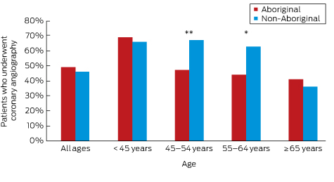

Aboriginal v non-Aboriginal patients: * P = 0.001; **P < 0.001 (logistic regression).

Box 4 –
Age-adjusted comparison of Aboriginal and non-Aboriginal patients with acute coronary syndromes who underwent coronary angiography, based on chart review data, according to guideline risk stratification
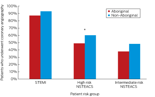

STEMI = ST-elevation myocardial infarction; NSTEACS = non-ST-elevation acute coronary syndrome. Aboriginal v non-Aboriginal patients: * P = 0.02 (logistic regression).

Box 5 –
Documentation of clinical and non-clinical factors in treating Aboriginal and non-Aboriginal patients with acute coronary syndromes without coronary angiography, chart review data

Aboriginal patients

Non-Aboriginal patients

Odds ratio for Aboriginal patients undergoing coronary angiography


Unadjusted (95% CI)

P

Age-adjusted (95% CI)

P


Number of patients

274

274

Coronary angiography not undertaken

139 (51%)

118 (43%)

1.4 (1.0–1.9)

0.07

1.6 (1.1–2.3)

0.01

Reason for not undertaking angiography

Patient-related factors

28 (20%)

8 (7%)

3.5 (1.5–8.4)

0.01

3.4 (1.4–8.2)

0.01

Patient decision

10

5

Discharge against medical advice

15

3

Known medication non-adherence

3

0

Clinical/medical factors

61 (44%)

98 (83%)

0.2 (0.1–0.3)

< 0.001

0.1 (0.1–0.3)

< 0.001

Angiography booked

7

10

Non-invasive test for ischaemia

11

41

Symptoms deemed non-ischaemic

22

15

Comorbidities/palliative care

7

14

Known anatomy, medical management

14

18

Unclear

50 (36%)

12 (10%)

5.3 (2.5–11)

< 0.001

5.7 (2.7–12.3)

< 0.001


Box 6 –
Guideline-recommended therapies at discharge for Aboriginal and non-Aboriginal patients with an acute myocardial infarction, chart review data

Discharge therapy

Aboriginal patients

Non-Aboriginal patients

Odds ratio for Aboriginal patients


Unadjusted (95% CI)

P

Age-adjusted (95% CI)

P


Number of patients*

134

150

Aspirin

119 (90%)

144 (95%)

0.5 (0.2–1.2)

0.12

0.5 (0.2–1.2)

0.11

Statin

118 (89%)

130 (93%)

0.6 (0.3–1.5)

0.32

0.6 (0.2–1.4)

0.24

β-Blocker

71 (54%)

67 (48%)

1.3 (0.8–2.1)

0.30

1.3 (0.8–2.0)

0.34

Angiotensin converting enzyme inhibitor or angiotensin receptor blocker

99 (76%)

113 (82%)

0.7 (0.4–1.2)

0.17

0.7 (0.4–1.3)

0.22

Cardiac rehabilitation referral

52 (50%)

54 (46%)

1.2 (0.7–2.0)

0.57

0.9 (0.5–1.6)

0.79

Ejection fraction assessed

66 (50%)

65 (44%)

1.3 (0.8–2.0)

0.33

1.2 (0.7–1.9)

0.57


Telehealth could deliver massive savings: CSIRO

Using telehealth technology to help the chronically ill to monitor and manage their condition at home could almost halve mortality rates and save the health budget up to $3 billion a year, according to CSIRO researchers.

Announcing the results of a 12-month trial, the CSIRO team reported that chronically ill patients provided with a telehealth service in their home not only had reduced mortality, but had less need for medical care and experienced shorter stays in hospital.

The outcomes add to evidence of the potential for telehealth technology to significantly improve the lives of patients while at the same time reducing the cost of their care.

The trial involved 287 patients with an average age of 71 years, who had at least one chronic illness such as congestive heart failure, chronic obstructive pulmonary disease, diabetes, hypertension and coronary heart disease and had been hospitalised twice in the preceding year.

They were each provided with an internet-connected telemedicine device that could monitor vital signs including ECG, heart rate, lung function, blood pressure, oxygen saturation, weight and temperature as well as video conferencing and messaging capabilities.

Patients were asked take their measurements once a day.

Participants reported benefits including the early detection of potentially deadly heart problems, a sharp fall in the number of visits to the doctor, and greater understanding of their illness and how to manage it.

Lead researcher Dr Rajiv Jayasena said these improvements resulted in a 24 per cent saving on Medicare costs for participants, as well as a 36 per cent reduction in hospital visits, a 42 per cent drop in the length of hospital stays and a 40 per cent decline in the mortality rate.

Telehealth Nurse Coordinator at Djerriwarrh Health Services, Lay Yean Woo said the system allowed her to monitor her patients and detect any abnormalities from her office, saving time that can be spent seeing more patients.

“This technology as helped me as a nurse and this has made my time more efficient in the way I deliver my service,” Ms Woo said. “Also, with the time that has been freed up, I can look at more new clients being referred to me. At the end of the day I know they are better looked after.”

While older Australians have some health habits – only 7 per cent smoke and 41 per cent report undertaking regular physical activity – 70 per cent are overweight or obese, almost a third consider their health is poor or only fair, and 20 per cent have problems that severely or profoundly limit their mobility.

As life expectancy has increased, more patients are developing chronic and complex health problems. Caring for them is placing an increasing demand on the health system, and the pressure is likely to intensify as their numbers swell. Currently, around 15 per cent of the population is 65 years or older, but the Australian Institute of Health and Welfare estimates that proportion will reach 22 per cent by the middle of the century and 24 per cent by 2096.

Dr Jayasena said that, with older patients with multiple chronic diseases accounting for 70 per cent of health spending, these benefits had the potential to deliver significant savings to the health budget.

The CSIRO has calculated that if the telehealth service was rolled out to the half a million Australians it considers would be good candidates, the nation could save up to $3 billion a year on health costs.

“Our research showed that the return on investment of a telemonitoring initiative on a national scale would be in the order of five to one by reducing demand on hospital inpatient and outpatient services, reduced visits to GPs, reduced visits from community nurses and an overall reduced demand on increasingly scarce clinical resources,” Dr Jayasena said.

The CSIRO, through its Smart Safer Homes initiative, is also fitting homes with sensors that track patient movement and raise the alarm when something out of the ordinary, such as being still on the ground for a period of time, happens.

Adrian Rollins

Patient charges rising fast

Patient out-of-pocket costs have surged and are now growing at their fastest pace in four years as general practices react to the financial squeeze from frozen Medicare rebates and rising running costs.

While the Federal Government has trumpeted official figures showing the proportion of GP services being bulk billed has risen to a record high of 85.1 per cent, the statistics also indicated that those patients that are being charged a fee are paying more.

Medicare data show that average out-of-pocket costs reached $34.25 last financial year, up 6.5 per cent from 2014-15 – the fastest pace of growth since 2011-12 and well above the rate of inflation.

The increase in patient charges follows warnings from AMA President Dr Michael Gannon that many general practices were “now at breaking point” because of the Medicare rebate freeze, cuts to incentive payments and reduced mental health funding.

“Many patients who are currently bulk billed will face out-of-pocket costs well over $20,” Dr Gannon said.

Related: Rebate freeze ‘must go’: Gannon

Hopes that the Turnbull Government, stung by voters over health policy, might move the scrap the rebate freeze are fading, heightening concerns that hard-pressed medical practices will have little choice but to abandon or cut back on bulk billing and increase charges for those patients judged to be able to pay a fee.

But instead, the Government has used the high incidence of bulk billing to argue its policies are sustainable.

Health Minister Sussan Ley seized on the increase in the bulk billing rate, claiming it was “good news for Australians”.

Ms Ley said the figures showed 123 million GP services were fully funded by the Government last financial year, and put the lie to Labor claims that the Government was anti-Medicare.

“These figure expose the blatant and remorseless Mediscare lies Labor have been telling the Australian public over the last 12 months,” Ms Ley said. “There’s no doubt we still have work to do, but Australians should tale assurance from the fact no Government has invested more into Medicare than the Turnbull Government.”

Related: Why doctors will stop bulk billing

But Shadow Health Minister Catherine King said the figures seized on by the Government were misleading because they focused solely the number of services that were bulk billed, rather than the number of patients, and ignored the rise in out-of-pocket costs.

Ms King said that as the rebate freeze has continued, a growing number of practices were abandoning bulk billing, including on Magnetic Island and in Hobart.

“Australians know that Malcolm Turnbull’s six-year freeze on Medicare rebates is driving bulk billing down and out-of-pocket costs up,” Ms King said. “The Government’s insistence otherwise only shows how out of touch they are.”

In his 17 August speech to the National Press Club, AMA President Dr Michael Gannon reiterated the AMA’s opposition to the rebate freeze, which he warned was undermining general practice, which was one of the key strengths of the nation’s health system.

“General practice has been under sustained pressure for years,” Dr Gannon said. “GPs have been treated poorly by both Coalition and Labor governments.”

The AMA President said that the ageing population and the growing burden of chronic and complex disease meant GPs were seeing more patients than ever before – an extra 42 million services in the past decade.

Despite this growth in demand, Government support for GPs was in decline.

“GPs are caught in a diabolical squeeze,” Dr Gannon said. “They are caring for increasingly sick patients while the Government tightens the financial screws in the name of budget repair.”

“GPs are now at breaking point. Many patients who are currently bulk billed will face out-of-pocket costs well over $20,” he warned.

Latest news: