×

Tick-borne infectious diseases in Australia

The incidence of tick-related medical problems in Australia is largely unknown. Appropriate diagnostic tests are not always available and, of all tick-related diseases, only Q fever is notifiable.1 Anecdotally, however, many patients present to their doctor after a tick bite. This narrative review focuses on tick-borne infections but also touches briefly on other medical problems caused by tick bites.

Australian ticks

There are many different species of ticks in Australia. Only a few species are known to bite humans, and the microbes within these particular ticks — viruses, bacteria or protozoa — are potential causes of infection in humans who are bitten (Box).

However, the mere detection of a potential human pathogen in a tick does not mean that it can be transmitted to a person when bitten. To be transmitted to a person, the microbe must be present in the salivary glands of the tick while it is feeding.

Most studies of Australian ticks to date have investigated the whole microbiome of the tick and not their salivary glands specifically. Some pathogens may be present in the tick faeces, but transmission would require the patient to scratch the faeces into their skin — an unlikely scenario in most cases.

To be confident that a microbe in a tick is responsible for a particular illness in a patient bitten by that tick, it is essential to also detect the microbe in the sick patient, either directly by culture or detection of microbial nucleic acid or antigen, or indirectly by detecting newly produced antibodies to the microbe in the patient’s blood. This requires the diagnostic laboratory to have assays that are both sensitive and specific for detecting the microbe in question. Few such assays are currently available in Australian diagnostic laboratories. Antibody assays are designed using antigens from a specific microbe and in many cases these microbes are not present in Australia. Such assays, even if reasonably sensitive and specific, must have acceptable positive and negative predictive values in Australia, so that patients and their doctors can have confidence in either a positive or negative result from any particular laboratory assay.

There have been two major studies of Australian ticks,2,3 which defined their variety and number. Australian ticks are divided into two large groups: soft-bodied and hard-bodied, comprising 14 and 56 species respectively.4 Five species of ticks have been introduced into Australia,4 but most are not important in biting humans. Of the endemic species, which are the majority, two are notorious for biting humans. On the east coast of Australia, the paralysis tick (Ixodes holocyclus) is the most notable; elsewhere in Australia, it is the ornate kangaroo tick (Amblyomma triguttatum). Both ticks are known to sometimes transmit human pathogens to people when they bite. The southern reptile tick (Bothriocroton hydrosauri) also bites humans and transmits infection (Box).3

There are four stages in the life history of ticks: egg, larva, nymph and adult (male and female). The larva, nymph and adult female (egg-producing) stages require a blood meal from a vertebrate animal to metamorphose into the next life stage. The stages that most often cause problems to humans are nymphs and adult females.

Most people are bitten by ticks without any problem arising from this abnormal host–ectoparasite feeding interaction. Humans are incidental hosts and attached ticks are usually detected by the individual within a few hours, or a day at the most, and killed. Most are probably not even aware when they are bitten by a tick, as the tick injects a local anaesthetic into the skin. However, once it starts to feed, it becomes noticeable as it enlarges.

The range of problems that may occur after tick bites can be classified as follows: infection; allergy; paralysis; autoimmunity; post-infection fatigue; and Australian multisystem disorder.

Viral infections

Although there are several viral infections associated with ticks in other parts of the world (eg, tick-borne encephalitis virus in Europe), there are no definite tick-borne viral infections of humans yet discovered in Australia. The seabird soft tick (Ornithodoros capensis) has been shown to contain several viruses including the Saumarez Reef virus.5 When this tick bites humans, a dermatological condition develops in some patients, but it is not yet clear whether this is due to a virus or an allergen in the tick saliva. A phlebovirus present in an Australian bird tick (I. eudyptidis) is pathogenic for the shy albatross6,7 and is closely related to the human pathogenic viruses that cause severe fever with thrombocytopenia syndrome and Heartland virus disease. This suggests that human pathogenic viruses may be present in Australian ticks, although there is currently no evidence of I. eudyptidis biting humans.

Bacterial infections

Many different bacteria have been detected in Australian tick species,813 mostly using molecular techniques. Some are known human pathogens or are closely related phylogenetically to known human pathogens; others are unique bacteria that are part of the tick microbiome.

Apart from the occasional local bacterial infection at the tick bite site (eschar), there are only two definite, known systemic infections following tick bites in Australia — rickettsial infection and Q fever — although there are other possible microbial pathogens and possibly as yet unknown infections (Box).

The two genera of bacteria currently confirmed as Australian tick-transmitted human pathogens are rickettsial species and Coxiella burnetii.

Rickettsial species

In Australia, rickettsial species cause Queensland tick typhus, Flinders Island spotted fever and Australian spotted fever. However, tick-transmitted rickettsiae in various parts of the world have recently been reviewed,14 and the findings emphasise the possibility that Australian clinicians may encounter patients who have returned to Australia after travelling and who present with a tick-borne infection contracted in another country.

The first case of Australian tick typhus was reported in 1946 from north Queensland15 and later that same year a series of cases, including the isolation of the causative agent, Rickettsia australis, from patients was described.16 The organism has since been isolated from a patient in south-east Queensland17 and from I. holocyclus and I. tasmani ticks.18 Queensland tick typhus was the first tick-transmitted infection recognised in Australia. It is seen regularly on the east coast of Australia from the Torres Strait Islands to the south-eastern corner of Victoria. The northern suburbs of Sydney are a particularly common location for transmission of this infection.19,20 Although often a mild condition involving fever, rash and eschar and readily treated with a short course of doxycycline, the infection may be severe21,22 or fatal,23 and may have unusual feaures.24 In north-eastern New South Wales, 15.4% of paralysis ticks contained R. australis.13 Hence, being bitten by this tick, in this location, appears to offer a 1 in 6 risk of being infected with the rickettsia.

A different rickettsial infection (Flinders Island spotted fever) has been observed in patients from Flinders Island, Tasmania.25,26 R honei was isolated from febrile patients27 and shown to be genetically different from R. australis. The tick vector was the southern reptile tick, which is known to bite humans, and on Flinders Island, 63% of these ticks were found to contain R. honei.28 Flinders Island spotted fever is now known to also occur in South Australia,29,30 Western Australia31 and other parts of the world.14

A related bacterium, Rickettsia honei subsp. marmionii, causes a similar infection, Australian spotted fever, and has been detected in the ticks I. tasmani (unpublished data) and Haemaphysalis novaeguineae in Queensland,32 and has been associated with several cases of human disease in eastern Australia.33

Two further species of rickettsia identified in Australian ticks may be considered potential human pathogens, although their presence in febrile patients is yet to be confirmed. Rickettsia gravesii has been detected in ornate kangaroo ticks in WA34 and Queensland (unpublished data). In a WA study, rogainers (outdoor recreationists) had a significantly higher seroprevalence to spotted fever group rickettsiae than controls with minimal bush exposure,35 suggesting exposure to a possible tick-transmitted rickettsia. A Tasmanian study found that 55% of I. tasmani ticks collected from Tasmanian devils contained rickettsial DNA. Further molecular characterisation of the DNA demonstrated sufficient divergence from previously described species to designate this new organism Candidatus Rickettsia tasmanensis.36 Because I. tasmani is known to bite humans, this rickettsia must be considered as a potential human pathogen.

Coxiella burnetii

This bacterium causes Q fever and usually infects humans by inhalation of infectious aerosols from carrier vertebrate animals such as goats, sheep, cattle and domestic pets. However, it is present in both paralysis ticks13 and ornate kangaroo ticks,37,38 and although, anecdotally, there are other cases of Q fever being transmitted by ticks, there is only one published case where the patient developed pericarditis, a rare presentation of Q fever, after being bitten by an ornate kangaroo tick.39

In north-eastern NSW, 5.6% of paralysis ticks contained the com1 gene of C. burnetii.13 This bacterium has been isolated from the bandicoot tick (Haemaphysalis humerosa) from both sides of Australia,40,41 although it is unlikely that this tick species bites humans.

Other possible bacterial pathogens causing rickettsial illness

Candidatus Neoehrlichia mikurensis has been shown to be a human pathogen in other countries,14 causing febrile illness and post-infection fatigue especially in immunocompromised patients. Recent Australian studies demonstrated the presence of Candidatus Neoehrlichia spp. in paralysis ticks,11,12 but their presence in Australian patients is yet to be shown.

Anaplasma and Ehrlichia species have been detected by molecular means in paralysis and ornate kangaroo ticks,11 and these bacteria from the ornate kangaroo tick in the southwest of WA have been named Anaplasma bovis strain Yanchep and Candidatus Ehrlichia occidentalis, respectively (personal communication, Alex Gofton, PhD student, Vector and Waterborne Pathogens Research Group, Murdoch University, January 2017). Certain species of these bacterial genera are known to be human pathogens (eg, E. chaffeensis, A. phagocytophilum). There is thus a possibility that these Australian bacteria may also be human pathogens.

Although a Borrelia species has been detected in the Australian echidna tick (Bothriocroton concolor),42 this bacterium belongs to a unique clade unrelated to the Borrelia species responsible for causing Lyme disease. This tick is not known to bite humans, so the bacterium is unlikely to be a human pathogen. A Borrelia species detected in native rats was not virulent for a human after experimental challenge.43 Lyme disease bacteria are probably not present in Australian ticks.10,44,45

Fancisella spp. are tick-transmitted bacteria that cause classic tularaemia. The tropical reptile tick from northern Australia (Amblyomma fimbriatum), which is not known to bite humans, has been shown to contain DNA from this bacterium.46 A case of a localised Francisella infection following a bite from a ring-tail possum has been reported,47 but it is not yet clear whether tularaemia is a tick-transmitted infection in Australia.

Protozoal infections

Babesia spp. are recognised human and animal pathogens transmitted by tick bites, especially in the northern hemisphere. In Australia, cattle are often infected with B. bigemina and/or B. bovis (cattle tick fever) via the bite of the Australian cattle tick (Rhipicephalus australis); and dogs with B. vogeli and/or B. gibsoni via the brown dog tick (R. sanguineus) and possibly the bush tick (Haemaphysalis longicornis).48,49 Only the bush tick is thought to bite humans.

A single case of human babesiosis caused by B. microti has been described in an Australian man who lived in close proximity to dogs but who did not recall being bitten by a tick and had not travelled outside of Australia for nearly 40 years.49 This was thought to have been a locally acquired infection, but there have been no subsequent cases of human babesiosis diagnosed in Australia.

Allergy following tick bites

A local allergic reaction to ticks is not uncommon and may take the form of urticaria or induration (due to tick saliva), scrub itch (due to infestations of nymphs) or rash.5052

Occasionally, the allergic reaction can be systemic, including wheezing, anaphylaxis and even death.53 Severe allergy has recently been described following prior sensitisation of a patient due to the ingestion of red meat.54

Paralysis following tick bites

I. holocyclus is known as the Australian paralysis tick because it injects a mixture of neurotoxins into its host when it bites. The role of these toxins for the tick is uncertain, but they often have a profound impact on the host animal. The toxins, known as holocyclotoxins, are small, cyclic polypeptides similar to botulinum toxin. They can affect native animals, family pets and occasionally humans, especially if they are small,55,56 and may cause ataxia followed by an ascending, symmetrical, flaccid paralysis similar to Guillain-Barré syndrome. Cranial nerves may be involved, leading to facial paralysis or ophthalmoplegia. The paralysis can extend even after the tick has been removed. There have been human deaths due to tick toxin, but not for many decades.55

Autoimmunity following tick bites

There is one report of Graves’ disease developing in a patient bitten by an unknown species of Australian tick in WA.57 The patient also had mild rickettsial infection following the bite. It was hypothesised that molecular homology between the thyroid-secreting hormone receptor of the patient and the rickettsial ATPase enzyme resulted in the synthesis of an antibody that cross-reacted with the host thyroid receptor, leading to increased synthesis of thyroid hormones.

Post-infection fatigue

This phenomenon is well known to be a consequence of several infections (eg, Ross River virus infection, Q fever, Epstein-Barr virus infection), although the antecedent infection may not be clearly identified by the patient. It is not yet widely recognised as a problem following rickettsial infection, although it has been suggested by a study involving two large cohorts of fatigued and non-fatigued patients,58 and a case report.59 In addition, there was a report of endemic typhus in SA,60 where patients often had a prolonged fatigue-like condition.

Australian multisystem disorder

This term has been proposed to describe patients with a range of symptoms of currently unknown aetiology, although they have been linked to tick bites in Australia.45 The main symptoms are fatigue, joint and muscle pain, and neurocognitive impairment, but vary from patient to patient. This is the group of patients who may have described themselves as having chronic Lyme (or Lyme-like) disease.44

Because so little is known about the medical effects of tick bites in Australia, it is important for medical practitioners to keep an open mind when dealing with patients who speak of problems associated with tick bites. While the patient may well have other underlying medical issues brought to light by the tick bite, a considered investigation of the whole clinical story is indicated.

If the tick bite is recent (eg, within 4 weeks) and the patient is symptomatic, an EDTA blood sample should be sent to a diagnostic laboratory for microbial polymerase chain reaction testing and culture, accompanied by a serum sample for antibody testing. This acute serum should be stored by the laboratory and tested in parallel with a later serum from the patient, looking for seroconversion or a significant rise in antibody titre, if the patient continues to be unwell and has not responded to treatment. The second (convalescent) serum is an important sample, as it may well contain antibodies to the causative microbe that were absent in the first (acute) serum.

However, when the tick bite has occurred some time ago (more than 8 weeks), serology is difficult to interpret, because antibody titres are stable and may reflect either recent or long-past infection.

In relation to Lyme disease, given the likely absence of the relevant bacteria in Australian ticks,10,44,45 there is little value in laboratory testing for the disease if the patient has not been to an endemic region of the world.

Conclusion

Much remains to be learned about the medical consequences of tick bites in Australia. While rickettsial infections are currently the most commonly known, it is likely that ongoing research will reveal new tick-borne viral, bacterial and protozoal infections, including the possibility of zoonotic transmission from wild and domestic mammals and birds which have been bitten by ticks.

This highlights the importance of the One Health concept (https://www.cdc.gov/onehealth), which recognises the importance of the interaction between human health, animal health and the environment, and will enable the identification of new and emerging diseases.

Box –
Australian tick species known to bite humans, and associated pathogens and diseases

Tick species

Common name

Known human pathogen

Disease

Possible human pathogen


Ixodes holocyclus

Paralysis tick (scrub tick in Queensland)

Rickettsia australis

Queensland tick typhus

Candidatus Neoehrlichia spp.

Coxiella burnetii

Q fever

Bartonella henselae; Ehrlichia spp.

Ixodes tasmani

Common marsupial tick

R. australis

Queensland tick typhus

Candidatus R. tasmanensis

R. honei subsp. marmionii

Australian spotted fever

Bartonella spp.

Ixodes cornuatus

Southern paralysis tick

R. australis

Queensland tick typhus

Amblyomma triguttatum

Ornate kangaroo tick

C. burnetii

Q fever

R. gravesii; Anaplasma sp.; Ehrlichia sp.

Bothriocroton hydrosauri

Southern reptile tick

R. honei

Flinders Island spotted fever

Haemaphysalis novaeguinae

No common name

R. honei subsp. marmionii

Australian spotted fever

Haemaphysalis longicornis

Bush tick (introduced, not native to Australia)

Babesia sp.

Ornithodoros capensis

Seabird soft tick

Virus


The Australasian Society for Infectious Diseases and Refugee Health Network of Australia recommendations for health assessment for people from refugee-like backgrounds: an abridged outline

There are currently more than 65 million people who have been forcibly displaced worldwide, including 21.3 million people with formal refugee status, over half of whom are aged under 18 years.1 More than 15 000 refugees have resettled in Australia in the 2015–16 financial year, which includes a proportion of the 12 000 refugees from Syria and Iraq recently added to Australia’s humanitarian intake.2 In addition, around 30 000 asylum seekers who arrived by plane or boat are currently in Australia awaiting visa outcomes.3

People from refugee-like backgrounds are likely to have experienced disruption of basic services, poverty, food insecurity, poor living conditions and prolonged uncertainty; they may have experienced significant human rights violations, trauma or torture. These circumstances place them at increased risk of complex physical and mental health conditions. They face numerous barriers to accessing health care after arrival in Australia, such as language, financial stress, competing priorities in the settlement period, and difficulties understanding and navigating the health care system.46 Most people require the assistance of an interpreter for clinical consultations.7 Offering a full health assessment to newly arrived refugees and asylum seekers is a positive step towards healthy settlement, and helps manage health inequity through the provision of catch-up immunisation and the identification and management of infectious and other health conditions.

These guidelines update the Australasian Society of Infectious Diseases (ASID) guidelines for the diagnosis, management and prevention of infectious diseases in recently arrived refugees8 published in 2009 and previously summarised in the MJA.9 When these recommendations were first published, more than 60% of humanitarian entrants arriving in Australia were from sub-Saharan Africa10 and had a high prevalence of malaria, schistosomiasis and hepatitis B virus (HBV) infection.1115 The initial guidelines were primarily intended to help specialists and general practitioners to diagnose, manage and prevent infectious diseases. Since then, there have been changes in refugee-source countries — with more arrivals from the Middle East and Asia and fewer from sub-Saharan Africa16,17 — and an increased number of asylum seekers arriving by boat,18 alongside complex and changing asylum seeker policies and changes in health service provision for these populations. In this context, we reviewed the 2009 recommendations to ensure relevance for a broad range of health professionals and to include advice on equitable access to health care, regardless of Medicare or visa status. The revised guidelines are intended for health care providers caring for people from refugee-like backgrounds, including GPs, refugee health nurses, refugee health specialists, infectious diseases physicians and other medical specialists.

This article summarises the full guidelines, which contain detailed literature reviews, recommendations on diagnosis and management along with explanations, supporting evidence and links to other resources. The full version is available at http://www.asid.net.au/documents/item/1225.

Methods

The guideline development process is summarised in Box 1. The two key organisations developing these guidelines are ASID and the Refugee Health Network of Australia. ASID is Australia’s peak body representing infectious diseases physicians, medical microbiologists and other experts in the fields of the prevention, diagnosis and treatment of human and animal infections. The Refugee Health Network is a multidisciplinary network of health professionals across Australia with expertise in refugee health.20

We defined clinical questions using the PIPOH framework (population, intervention, professionals, outcomes and health care setting).21 The chapter authors and the Expert Advisory Group developed recommendations based on reviews of available evidence, using systematic reviews where possible. Australian prevalence data also informed screening recommendations; for example, the low reported prevalence of chlamydia (0.8–2.0%) infections and absence of gonorrhoea infections in refugee cohorts in Australia13,2224 (and in other developed countries2527) informed the new recommendation for risk-based sexually transmitted infection (STI) screening.

Despite the intention to assign levels of evidence to each recommendation, there was limited published high level evidence in most areas, and virtually all recommendations are based on expert consensus. Consensus was not reached regarding the recommendations relating to human immunodeficiency virus (HIV) and STIs.

The term “refugee-like” is used to describe people who are refugees under the United Nations Refugee Convention,28 those who hold a humanitarian visa, people from refugee-like backgrounds who have entered under other migration streams, and people seeking asylum in Australia. “Refugee-like” acknowledges that people may have had refugee experience in their countries of origin or transit, but do not have formal refugee status.

Current pre-departure screening

All permanent migrants to Australia have a pre-migration immigration medical examination 3–12 months before departure,29 which includes a full medical history and examination. Investigations depend on age, risk factors and visa type,30 and include:

  • a chest x-ray for current or previous tuberculosis ([TB]; age ≥ 11 years);

  • screening for latent TB infection with an interferon-γ release assay or tuberculin skin test (for children aged 2–10 years, if they hold humanitarian visas, come from high prevalence countries or have had prior household contact);

  • HIV serology (age ≥ 15 years, unaccompanied minors);

  • hepatitis B surface antigen (HBsAg) testing (pregnant women, unaccompanied minors, onshore protection visas, health care workers);

  • hepatitis C virus (HCV) antibody testing (onshore protection visas, health care workers); and

  • syphilis serology (age ≥ 15 years, humanitarian visas, onshore protection visas).

Humanitarian entrants are also offered a voluntary pre-departure health check depending on departure location and visa subtype.31 The pre-departure health check includes a rapid diagnostic test and treatment for malaria in endemic areas; empirical treatment for helminth infections with a single dose of albendazole; measles, mumps and rubella vaccination; and yellow fever and polio vaccination where relevant. The current cohort of refugees arriving from Syria will have extended screening incorporating the immigration medical examination and pre-departure health check, with additional mental health review and immunisations.

People seeking asylum who arrived by boat have generally had a health assessment on arrival in immigration detention — although clinical experience suggests that investigations and detention health care varies, especially for children. However, asylum seekers who arrived by plane will not have had a pre-departure immigration medical examination.

General recommendations

Our overarching recommendation is to offer all people from refugee-like backgrounds, including children, a comprehensive health assessment and management plan, ideally within 1 month of arrival in Australia. This assessment can be offered at any time after arrival if the initial contact with a GP or clinic is delayed, and should also be offered to asylum seekers after release from detention. Humanitarian entrants who have been in Australia for less than 12 months are eligible for a GP Medicare-rebatable health assessment. Such assessments may take place in a primary care setting or in a multidisciplinary refugee health clinic. Documented overseas screening and immunisations, and clinical assessment should also guide diagnostic testing.

Health care providers should adhere to the principles of person-centred care when completing post-arrival assessments.32,33 These include: respect for the patient’s values, preferences and needs; coordination and integration of care with the patient’s family and other health care providers; optimising communication and education, provision of interpreters where required (the Doctors Priority Line for the federal government-funded Translating and Interpreting Service is 1300 131 450) and use of visual and written aids and teach-back techniques to support health literacy.34 It is important to explain that a health assessment is voluntary and results will not affect visa status or asylum claims.

Specific recommendations

Recommendations are divided into two sections: infectious and non-infectious conditions. Box 2 provides a checklist of all recommended tests, and Box 3 sets out details of country-specific recommendations. A brief overview is provided below. For more detailed recommendations regarding management, follow-up and considerations for children and in pregnancy, see the full guidelines.

Infectious conditions

TB:

  • Offer latent TB infection testing with the intention to offer preventive treatment and follow-up.

  • Offer screening for latent TB infection to all people aged ≤ 35 years.

  • Children aged 2–10 years may have been screened for latent TB infection as part of their pre-departure screening.

  • Screening and preventive treatment for latent TB infection in people > 35 years will depend on individual risk factors and jurisdictional requirements in the particular state or territory.

  • Use either a tuberculin skin test or interferon-γ release assay (blood) to screen for latent TB infection.

  • A tuberculin skin test is preferred over interferon-γ release assay for children < 5 years of age.

  • Refer patients with positive tuberculin skin test or interferon-γ release assay results to specialist tuberculosis services for assessment and exclusion of active TB and consideration of treatment for latent TB infection.

  • Refer any individuals with suspected active TB to specialist services, regardless of screening test results.

Malaria:

  • Investigations for malaria should be performed for anyone who has travelled from or through an endemic malaria area (Box 3), within 3 months of arrival if asymptomatic, or any time in the first 12 months if there is fever (regardless of pre-departure malaria testing or treatment).

  • Test with both thick and thin blood films and an antigen-based rapid diagnostic test.

  • All people with malaria should be treated by, or in consultation with, a specialist infectious diseases service.

HIV:

  • Offer HIV testing to all people aged ≥ 15 years and all unaccompanied or separated minors, as prior negative tests do not exclude the possibility of subsequent acquisition of HIV (note that consensus was not reached regarding this recommendation).

HBV:

  • Offer testing for HBV infection to all, unless it has been completed as part of the immigration medical examination.

  • A complete HBV assessment includes HBsAg, HB surface antibody and HB core antibody testing.

  • If the HBsAg test result is positive, further assessment and follow-up with clinical assessment, abdominal ultrasound and blood tests are required.

HCV:

  • Offer testing for HCV to people if they have:

    • risk factors for HCV;

    • lived in a country with a high prevalence (> 3%) of HCV (Box 3); or

    • an uncertain history of travel or risk factors.

  • Initial testing is with an HCV antibody test. If the result is positive, request an HCV RNA test.

  • If the HCV RNA test result is positive, refer to a doctor accredited to treat HCV for further assessment.

Schistosomiasis:

  • Offer blood testing for Schistosoma serology if people have lived in or travelled through endemic countries (Box 3).

  • If serology is negative, no follow-up is required.

  • If serology is positive or equivocal:

    • treat with praziquantel in two doses of 20 mg/kg, 4 hours apart, orally; and

    • perform stool microscopy for ova, urine dipstick for haematuria, and end-urine microscopy for ova if there is haematuria.

  • If ova are seen in urine or stool, evaluate further for end-organ disease.

Strongyloidiasis:

  • Offer blood testing for Strongyloides stercoralis serology to all.

  • If serology is positive or equivocal:

    • check for eosinophilia and perform stool microscopy for ova, cysts and parasites; and

    • treat with ivermectin 200 μg/kg (weight ≥ 15 kg), on days 1 and 14 and repeat eosinophil count and stool sample if abnormal.

  • Refer pregnant women or children < 15 kg for specialist management.

Intestinal parasites:

  • Check full blood examination for eosinophilia.

  • If pre-departure albendazole therapy is documented:

    • if there are no eosinophilia and no symptoms, no investigation or treatment is required; and

    • if there is eosinophilia, perform stool microscopy for ova, cysts and parasites, followed by directed treatment.

  • If no documented pre-departure albendazole therapy, depending on local resources and practices, there are two acceptable options:

    • empirical single dose albendazole therapy (age > 6 months, weight < 10 kg, dose 200 mg; weight ≥ 10 kg, dose 400 mg; avoid in pregnancy, class D drug); or

    • perform stool microscopy for ova, cysts and parasites, followed by directed treatment.

Helicobacter pylori:

  • Routine screening for H. pylori infection is not recommended.

  • Screen with either stool antigen or breath test in adults from high risk groups (family history of gastric cancer, symptoms and signs of peptic ulcer disease, or dyspepsia).

  • Children with chronic abdominal pain or anorexia should have other common causes of their symptoms considered in addition to H. pylori infection.

  • Treat all those with a positive test (see the full guidelines for details, tables 1.5 and 9.1).

STIs:

  • Offer an STI screen to people with a risk factor for acquiring an STI or on request. Universal post-arrival screening for STIs for people from refugee-like backgrounds is not supported by current evidence.

  • A complete STI screen includes a self-collected vaginal swab or first pass urine nucleic acid amplification test and consideration of throat and rectal swabs for chlamydia and gonorrhoea, and serology for syphilis, HIV and HBV.

  • Syphilis serology should be offered to unaccompanied and separated children < 15 years.

Skin conditions:

  • The skin should be examined as part of the initial physical examination.

  • Differential diagnoses will depend on the area of origin (see table 11.1 in full guidelines for details).

Immunisation:

  • Provide catch-up immunisation so that people of refugee background are immunised equivalent to an Australian-born person of the same age.

  • In the absence of written immunisation documentation, full catch-up immunisation is recommended.

  • Varicella serology is recommended for people aged ≥ 14 years if there is no history of natural infection.

  • Rubella serology should be completed in women of childbearing age.

Non-infectious conditions

Anaemia and other nutritional problems:

  • Offer full blood examination screening for anaemia and other blood conditions to all.

  • Offer screening for iron deficiency with serum ferritin to children, women of childbearing age, and men who have risk factors.

  • Check vitamin D status as part of initial health screening in people with one or more risk factors for low vitamin D.

  • People with low vitamin D should be treated to restore their levels to the normal range with either daily dosing or high dose therapy, paired with advice about sun exposure.

  • Consider screening for vitamin B12 deficiency in people with history of restricted food access, especially those from Bhutan, Afghanistan, Iran and the Horn of Africa.

Chronic non-communicable diseases in adults:

  • Offer screening for non-communicable diseases in line with the Royal Australian College of General Practitioners Red Book35 recommendations, including assessment for:

    • smoking, nutrition, alcohol and physical activity;

    • obesity, diabetes, hypertension, cardiovascular disease, chronic obstructive pulmonary disease and lipid disorders; and

    • breast, bowel and cervical cancer.

  • Assess diabetes and cardiovascular disease risk earlier for those from regions with a higher prevalence of non-communicable diseases, or those with an increased body mass index or waist circumference.

Mental health:

  • A trauma informed assessment of emotional wellbeing and mental health is part of post-arrival screening. Being aware of the potential for past trauma and impact on wellbeing is essential, although it is generally not advisable to ask specifically about details in the first visits.

  • Consider functional impairment, behavioural difficulties and developmental progress as well as mental health symptoms when assessing children.

Hearing, vision and oral health:

  • A clinical assessment of hearing, visual acuity and dental health should be part of primary care health screening.

Women’s health:

  • Offer women standard preventive screening, taking into account individual risk factors for chronic diseases and bowel, breast and cervical cancer.

  • Consider pregnancy and breastfeeding and offer appropriate life stage advice and education, such as contraceptive advice where needed, to all women, including adolescents.

  • Practitioners should be aware of clinical problems, terminology and legislation related to female genital mutilation or cutting and forced marriage.

Box 1 –
Guideline development process


  • An EAG, consisting of refugee health professionals, was formed and it included two ID physicians, an ID and general physician, two GPs, a public health physician, a general paediatrician and a refugee health nurse. An editorial subgroup was also formed.
  • The EAG determined the list of priority conditions in consultation with refugee health specialists and RACGP Refugee Health Special Interest Group clinicians, incorporating information from consultations with refugee background communities19 and previous ASID refugee health guidelines.
  • Each condition was assigned to a primary specialist author with paediatrician and primary care or specialist co-authors. Twenty-eight authors from six states and territories were involved in writing the first draft.
  • The EAG reviewed the first draft to ensure consistency with the framework and the rest of the guidelines. They were then revised by the primary authors.
  • External expert review authors reviewed the second draft and they were then revised by the primary authors.
  • The EAG and the refugee health nurse subcommittee reviewed the third draft.
  • The stakeholders reviewed the fourth draft: ASID, NTAC, RHeaNA, RACGP Refugee Health Special Interest Group, RACP, RACP AChSHM, the Victorian Foundation for the Survivors of Torture, the Multicultural Centre for Women’s Health, the Asylum Seeker Resource Centre, the Ethnic Communities Council of Victoria and community members.
  • The comments from the stakeholders were returned to the authors for review and the EAG compiled the final version.
  • ASID, RACP, NTAC and AChSHM endorsed the final version.

AChSHM = Australasian Chapter of Sexual Health Medicine. ASID = Australasian Society for Infectious Diseases. EAG = Expert Advisory Group. GP = general practitioner. ID = infectious diseases. NTAC = National Tuberculosis Advisory Council. RACGP = Royal Australian College of General Practitioners. RACP = Royal Australasian College of Physicians. RHeaNA = Refugee Health Network of Australia. Adapted from the ASID and RHeaNA Recommendations for comprehensive post-arrival health assessment for people from refugee-like backgrounds (2016; https://www.asid.net.au/documents/item/1225) with permission from ASID.

Box 2 –
Short checklist of recommendations for post-arrival health assessment of people from refugee-like backgrounds

Offer test to

Test

Comments and target condition


All

Full blood examination

Anaemia, iron deficiency, eosinophilia

Hepatitis B serology (HBsAg, HBsAb, HBcAb)

HBsAg testing introduced overseas in 2016 for Syrian and Iraqi refugee cohort and may have been completed in other groups

Strongyloides stercoralis serology

Strongyloidiasis

HIV serology*

≥ 15 years or unaccompanied or separated minor
Also part of IME for age ≥ 15 years

TST or IGRA

Offer test if intention to treat. All ≤ 35years; if≥ 35 years, depends on risk factors and local jurisdiction. TST preferred for children < 5 yearsTST or IGRA testing introduced in 2016 as part of IME for children 2–10 years (humanitarian entrants, high prevalence countries, prior household contact)
LTBI

Varicella serology

≥ 14 years if no known history of disease
Determine immunisation status

Visual acuity

Vision status, other eye disease

Glaucoma assessment

Africans > 40 years and others > 50 years

Dental review

Caries, periodontal disease, other oral health issues

Hearing review

Hearing impairment

Social and emotional wellbeing and mental health

Mental illness, trauma exposure, protective factors

Developmental delay or learning concerns

Children and adolescents
Developmental issues, disability, trauma exposure

Preventive health as per RACGP35

Non-communicable diseases, consider screening earlier than usual age

Catch-up vaccinations

Vaccine preventable diseases, including hepatitis B

Risk-based

Rubella IgG

Women of childbearing age
Determines immunisation status

Ferritin

Men who have risk factors, women and childrenIron deficiency anaemia

Vitamin D, also check calcium, phosphate, and alkaline phosphatase in children

Risk factors if dark skin or lack of sun exposure
Low vitamin D, rickets

Vitamin B12

Arrival < 6 months, food insecurity, vegan diet or from Bhutan, Afghanistan, Iran or Horn of Africa
Nutritional deficiency, risk for developmental disability in infants

First pass urine or self-obtained vaginal swabs for gonorrhoea and chlamydia PCR

Risk factors for STI or on request*

Syphilis serology

Risk factors for STIs, unaccompanied or separated minors. Part of IME in humanitarian entrants aged ≥ 15 years

Helicobacter pylori stool antigen or breath test

Gastritis, peptic ulcer disease, family history of gastric cancer, dyspepsia

Stool microscopy (ova, cysts and parasites)

If no documented pre-departure albendazole or persisting eosinophilia despite albendazoleIntestinal parasites

Country-based (Box 3)

Schistosoma serology

Schistosomiasis

Malaria thick and thin films and rapid diagnostic test

Malaria

HCV Ab, and HCV RNA if HCV Ab positive

HCV, also test if risk factors, regardless of country of origin


HBcAb = hepatitis B core antibody. HBsAb = hepatitis B surface antibody. HBsAg = hepatitis B surface antigen. HCV = hepatitis C virus. HCV Ab = hepatitis C antibody. HIV = human immunodeficiency virus. IGRA = interferon-γ release assay. IME = immigration medical examination. LBTI = latent tuberculosis infection. PCR = polymerase chain reaction. TST = tuberculin skin test. * The panel did not reach consensus on these recommendations. See full guideline at http://www.asid.net.au/documents/item/1225 for details.

Box 3 –
Top 20 countries of origin for refugees and asylum seekers2,3,16 and country-specific recommendations for malaria, schistosomiasis and hepatitis C screening*

Country of birth

Malaria36

Schistosomiasis37

Hepatitis C38


Afghanistan

No

No

No

Bangladesh

Yes

No

No

Bhutan

Yes

No

No

Burma

Yes

Yes

No

China

No

No

No

Congo

Yes

Yes

Yes

Egypt

No

Yes

Yes

Eritrea

Yes

Yes

No

India

Yes

Yes

No

Iran

No

No

No

Iraq

No

Yes

Yes

Lebanon

No

No

No

Pakistan

Yes

No

Yes

Somalia

Yes

Yes

No

Sri Lanka

Yes

No

No

Stateless

Yes

Yes

No

Sudan

Yes

Yes

No

Syria

No

Yes

Consider

Vietnam

No

No

No


* There are regional variations in the prevalence of these conditions within some countries. We have taken the conservative approach of recommending screening for all people from an endemic country rather than basing the recommendation on exact place of residence. Note that some refugees and asylum seekers may have been exposed during transit through countries not listed here. See full guideline for further details. † People with risk factors for hepatitis C should be tested regardless of country of origin. ‡ “Stateless” in this table refers to people of Rohingyan origin. Adapted from the ASID and RHeaNA Recommendations for comprehensive post-arrival health assessment for people from refugee-like backgrounds (2016; https://www.asid.net.au/documents/item/1225) with permission from ASID.

Translating our microbiome into medicine

Integrating contemporary microbiology with new sequencing technologies will allow us to better understand our microbiome and its relationships with health and disease

We live in a microbial world. We are surrounded by and in contact with microbes that support many natural and managed processes, from carbon capture by microalgae to support marine food webs, to the fermentations that produce beverages and foods that we consume and enjoy. Nor is our body a sterile environment, either inside or out. While it has long been recognised that animals, including humans, are colonised soon after birth (or hatching), changes in our perceptions of the human microbiota have arisen over the past decade via the step advances made in DNA and RNA amplification methods, sequencing technologies and computational biology — what has been referred as the “omics” era of research. All these approaches support an assessment of the microbial world without first having to culture microbes in a laboratory setting, and the capacity to characterise both the structure and function of entire microbial communities from real-world samples.

During the past decade, initiatives in North America, Europe and China have been at the forefront of providing a genomic-based characterisation of our microbiota — this is generically referred to as our microbiome, with the term “microbiota” restricted to the viable state.1 If our body is considered as a landscape, then it possess multiple environments with variations in temperature, water content, pH, exposure to light and gases, and oxygen availability. Despite these variations, microbiome profiling studies using 16S ribosomal RNA gene sequencing2 have shown that our recruitment of the microbial world to reside and interact with us is rather selective, constrained to around 10 of the 50 or so phyla that are currently known to exist on our planet, and with each site possessing its own signature profile of members from this broadest taxonomic classification.3 Evidence also now suggests that our first encounter with the microbial world occurs in utero via a placental microbiome,4 and other body sites and organs once considered to be sterile under healthy conditions, such as the lung, are now considered to possess their own community of commensal microorganisms.5 Even body sites such as the oral cavity and large bowel, which have long been studied using classical microbiology techniques, have been transformed in terms of our understanding of their form and function. We have learned that our microbiota ages with us, and can be shaped in a deterministic way by our own genetics and dietary pattern.6 More recently, time and cost constraints have lessened, allowing metagenomic sequencing,2 which provides an inventory of the complete genetic potential inherent to microbial communities, and may soon supplant microbiota profiling approaches. Both approaches have further expanded our awareness of the scope of the nutritional, structural and physiological impacts that the gut microbiota may have on host metabolism, (immuno)physiology and other homeostatic processes. Indeed, observational, cross-sectional and case–control studies have now been published reporting changes in the microbiome that are associated with various medical conditions. This has led to the use of the term “dysbiosis” to refer collectively to what is considered an aberrant microbial profile present with these conditions when compared with healthy subjects.7

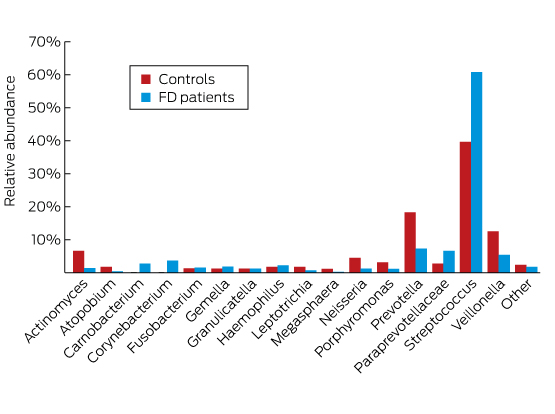
In a study of the duodenal mucosal microbiome, patients with functional dyspepsia were found to have a greater relative abundance of Streptococcus and decreases in the relative abundance of other genera such as Prevotella, Veillonella and Actinomyces compared with control subjects, suggesting that their symptoms may be related to alterations of their microbiome at this site (Box).8

It has also long been recognised that some animal models of disease, when maintained in a germ-free state, remain free of the phenotype. For example, germ-free animal models of inflammatory bowel disease remain quiescent until microbiota are introduced, which results in the rapid onset of disease.9 Conceptually similar observations have recently been made with respect to the role of the gut microbiota in trimethylamine formation from choline-rich diets and cardiovascular disease pathogenesis.10 Gnotobiotic mouse models are also now being used to validate that some specific commensal gut bacteria have protective effects, by positively affecting gut homoeostasis and attenuating inflammation or disease. For instance, Miquel and colleagues11 used gnotobiotic mice (Escherichia coli ± Faecalibacterium prausnitzii) to show that if F. prausnitzii is maintained at high levels in colitic mice, the disruption of the colonic epithelium is greatly attenuated.

Additionally, microbiota transplants in mice, using faecal slurries prepared from humans or from mice with specific phenotypes, have become a popular route for establishing that the microbiota, as a community, can elicit phenotypic changes in the host, from obesity12 to depressive-like behaviours.13 Such findings, along with the now well established clinical efficacy of faecal microbiota transplants (FMT) for alleviating Clostridium difficile infections, have led to a resurgent interest in this approach in Australia14 and abroad to treat a variety of medical conditions where gut dysbiosis is implicated.15,16 However, the current optimism for the use of FMT to treat a broader range of digestive diseases is moderated by our current understanding of the microbiome from both a donor and recipient context. While the donor’s stool microbiome (or its preparation) is widely accepted as a key influence on the therapeutic effect, it is also possible that patients could be exposed to additional risks transmitted via a donor’s stool. The resilience of the recipient’s microbiome to perturbations like FMT, and its recovery from such a challenge, will also likely affect efficacy. In that context, our gut microbiota is considered to have developed resilience relatively early in life,6 but our understanding of the concept of resilience and strategies that might support our management of it in a clinical setting are largely unexplored. Interestingly, a recent report of vaginal microbiota transfer from mothers to their infants delivered by caesarean section suggests this intervention, at a time when the human microbiota is most dynamic and pliable, might promote the development of a skin and oral microbiota more similar to that of their mothers compared with untreated infants.17 In summation, while the findings above represent some of the many findings linking our microbiota with our health, and reveal much promise and optimism for the use of our microbiota for future treatments, a combination of caution and refinement of approaches remains vital from an ethical, legal and social context. To this end, efforts such as the European consensus conference on FMT in clinical practice18 seek to promote an evidence-based approach as guidance for interested physicians.

In relation to key knowledge gaps that constrain the translation of our microbiome into medicine, metagenomic analysis of stool microbiota from various origins (Europe, North America, and Japan) suggests that these communities are similar but not universal.19 To date, equivalent datasets have not been produced more broadly across the Asia–Pacific region, despite the region’s genetically diverse populations with vastly different diets and lifestyles compared with those of the Americas, Europe, Africa and China. Common to these ethnic groups are the burdens of many, but not all, chronic Western immune-mediated and metabolic diseases and cancers, and at rapidly increasing incidence rates, suggesting a non-genetic basis for their penetration into these communities. Moreover, while the omics-based approaches have produced substantial advances in our knowledge of the human microbiome, much remains as “dark matter” in the form of unclassified genes or metagenomic species assembled from the data, including viruses, but for which no microbial isolate or biochemical characterisation is available.20 Indeed, we contend that there has been, to some extent, the development of a culture gap, both literally and figuratively, between the genomics-driven approaches and the other research elements inherent to environmental microbiology, which needs to be corrected. In this context, the lower eukaryotes (protozoa, yeasts and fungi) have been somewhat neglected during the omics era — in particular their roles as members of the gut microbiota21 — and warrant further investigation. It should also be noted that most research to date has focused on the stool microbiome. However, colonisation of the mucosa by specific bacteria (the mucosa-associated microbiome) and/or the study of microbiomes resident within other surfaces of the human body might be more relevant with respect to other medical conditions afflicting different body sites and organs.

In conclusion, exploring the microbiome and, in particular, the interactions between the microbiome and the human host, may substantially add to our knowledge of the pathophysiology of many diseases. While targeting the microbiome may offer new approaches to prevent or treat a broad spectrum of diseases, the interactions between host and the microbiome are complex. Simple but crude measures such as exposing patients to stool preparations (FMT) from healthy subjects might be attractive, but in the long term are unlikely to be the way forward, considering safety aspects and the need for standardised and contamination-free microbiota preparations that comply with good manufacturing practice standards. On the other hand, modulation of the microbiome with currently available probiotics — although more likely to be compliant with relevant manufacturing standards — may not deliver the potential benefits. Omics-based approaches will be crucial to revealing potential roles for the microbiome in health and disease, and when combined with the principles of environmental microbiology, including culture, offer the opportunity to shine a light on our microbial dark matter and refine the concepts of probiotics, prebiotics and FMT. The continued support and inspiration of the medical community will therefore be key for catalysing the translation of our new understanding of the microbiome into medicine.

Box –
Difference in the duodenal microbiome of patients with functional dyspepsia (FD) compared with controls, using 16S ribosomal RNA gene sequencing

Adapted with permission from Zhong et al.8

Australasian Society for Infectious Diseases: low value interventions

The challenge will be changing clinicians’ behaviour and practice so that the use of low value interventions decreases

In March 2015, the Australasian Society for Infectious Diseases (ASID) was one of 41 medical societies of the Royal Australasian College of Physicians to participate in the EVOLVE initiative, aimed at identifying five practices or interventions that were of low value or of limited usefulness.1 ASID members, including paediatricians, were surveyed and asked to short-list (and rank) suggested low value interventions (LVIs). From this survey emerged an overall short list that was circulated to respondents for further comment, and the final five LVIs were submitted to the ASID Council for endorsement.1

We present these five interventions below, with rationales as to why they are considered to be of low value. There are, of course, some uncommon situations where these interventions may demonstrate utility and we give some examples of these exceptions.

It is notable that four of the five interventions relate to the inappropriate use of antibiotics. Antibiotic use, both appropriate and inappropriate, is the major driver of antimicrobial resistance, which a recent World Health Organization report has highlighted as “an increasingly serious threat to global public health”.2 Inappropriate antibiotic use is also associated with a risk of Clostridium difficile infection,3 an unnecessary risk of developing antibiotic allergy and unjustified health care costs.

The five low value interventions

1. Prescribing antibiotics for asymptomatic bacteriuria

Asymptomatic bacteriuria (with or without pyuria) is common, particularly in older patients, and does not require treatment. Antibiotic treatment for asymptomatic bacteriuria does not decrease the incidence of symptomatic urinary tract infection or systemic sepsis. This also applies to patients with indwelling catheters: bacteriuria is almost universal in patients with urinary catheters in situ for more than a few days, and antimicrobial therapy does not decrease their risk of clinical symptoms or sepsis.

Thus, it is generally recommended that clinicians request urine samples for microscopy and culture only when patients have symptoms. Because a positive urine culture from an asymptomatic patient may trigger a decision to prescribe unnecessary antibiotic therapy, not ordering the test is the best way to avoid this situation. There are a few situations where antibiotics are indicated for asymptomatic bacteriuria. The most common are during pregnancy,4 when screening should be performed at the first antenatal visit,3 and preoperatively for patients undergoing a urological procedure in which mucosal bleeding is anticipated.4

2. Taking a swab of a leg ulcer without signs of clinical infection and treating the patient with antibiotics against the identified bacteria

Leg ulcers, such as venous ulcers, should not be investigated or treated for bacterial infection in the absence of clinical evidence of infection, such as purulent discharge or spreading erythema. There is no evidence that antibiotic therapy promotes wound healing in this setting5 Swabbing an ulcer and performing microscopy and culture in the absence of clinical signs of infection may identify commensal flora of no clinical relevance. Even if a potential pathogen such as Staphylococcus aureus or a β-haemolytic streptococcus is present, antimicrobial therapy in the absence of significant inflammation is not recommended. These recommendations for leg ulcers (not to take a swab or treat with antibiotics unless there are clinical symptoms of infection) apply to many other skin conditions that may present with leg erythema, such as lower leg venous stasis, contact dermatitis, arterial ischaemia and dependent oedema.

3. Treating upper respiratory tract infections with antibiotics

Most uncomplicated upper respiratory tract infections (URTIs) are viral in aetiology and antibiotic therapy is not indicated. This is particularly relevant in young children, who frequently receive unnecessary antibiotic therapy for URTIs. The antibiotic volume of the Australian Therapeutic Guidelines recommends avoiding “routine use” of antibiotic therapy for acute rhinosinusitis.3 Antibiotics are frequently prescribed for a purulent nasal discharge or to prevent secondary bacterial pneumonia,6 but there is no evidence to support such use.

Symptomatic management and education about the lack of benefit and potential adverse effects of antibiotics are key in this setting. Education can change doctors’ behaviour with regard to inappropriate prescribing of antibiotics,7 and education for patients and their parents or caregivers should help them to understand that improvement in the patient’s condition came with time and not as a result of inappropriately prescribed antibiotics.

There are specific URTIs where antibiotics are indicated, and these include Streptococcus pyogenes pharyngitis and Bordetella pertussis infection.

4. Investigation for faecal pathogens in the absence of diarrhoea or other gastrointestinal symptoms

Microscopy and culture or, more recently (and particularly), multiplex polymerase chain reaction (PCR) testing of faeces, should not be performed in the absence of diarrhoea or other gastrointestinal symptoms. Microbiology laboratories should not process a formed faecal specimen. Moreover, antimicrobial treatment for a potential gastrointestinal pathogen is not indicated in the absence of symptoms. For example, a patient whose diarrhoea has resolved by the time a microbiological diagnosis of C. difficile infection is made does not require treatment.

The recent introduction of faecal multiplex DNA-based diagnostic (PCR) methods has resulted in increased detection and reporting of several rarely pathogenic protozoa, especially Blastocystis hominis and Dientamoeba fragilis, as molecular methods are considerably more sensitive than microscopy. These organisms are often found in patients who are asymptomatic or whose symptoms are incompatible with enteric infection. Antimicrobial treatment is generally unnecessary and not recommended. The Australian and New Zealand Paediatric Infectious Diseases Group has highlighted this issue8 and, following consultation, the Royal College of Pathologists of Australasia now recommends that diagnostic laboratories use multiplex PCR tests without targets for these two protozoans.9

There are times where testing of non-diarrhoea stool may be indicated. These include:

  • screening of refugees for chronic parasitic infection that may be asymptomatic (eg, schistosomiasis and strongyloidosis);10

  • neurological syndromes (eg, acute flaccid paralysis) where enteroviruses may be implicated on epidemiological grounds;11 and

  • to confirm faecal clearance of Salmonella typhi or Salmonella paratyphi after treatment of enteric fever in food handlers, under the direction of public health authorities.

5. Ordering multiple serological investigations for patients with fatigue without a clinical indication or relevant epidemiology

It is very unusual for serological testing (eg, for brucellosis, Q fever, rickettsial disease, syphilis) to identify an underlying cause of fatigue if there is no clinical indication of an infectious cause on history or examination and in the absence of relevant epidemiology (ie, known risk factors).12 This is especially true if the patient has been fatigued for a prolonged period.

Acute (IgM) serological testing is notoriously non-specific and often leads to further unnecessary investigations and treatments, with potential adverse effects, inconvenience, erroneous diagnoses (eg, in the case of false positive results) and cost.

Use of low value interventions

Although there are no national data on how often the LVIs described above are used in current clinical practice, some studies suggest they are likely to be widespread. In one report from New Zealand, more than three-quarters of patients with an URTI received antibiotics.13

The underlying reasons for the popularity of these interventions are multiple and include: lack of an evidence base for treating some conditions; the expectations of patients and caregivers;14 suboptimal training and work pressure for clinicians;15 the anxiety of missing the diagnosis of a significant condition;16 and fear of litigation.15 Broad spectrum testing and therapy may be perceived (almost always erroneously) to compensate in some way for the lack of an evidence base.14

The EVOLVE initiative continues to be a useful vehicle to question common but non-evidence-based and potentially wasteful and harmful clinical practices, and to identify and discuss interventions that are of low value. However, the lack of usefulness of many of these LVIs is already well known, so it is important to question why they are still being used.

The challenge for ASID, and for all the societies involved in the EVOLVE initiative, is influencing behaviour to change practice so that the use of identified LVIs by medical practitioners decreases. Widespread and ongoing education, directed both at practitioners and the community, should be enhanced. ASID’s participation in the expert working groups that develop the antibiotic volume of the Australian Therapeutic Guidelines3 is likely to influence inappropriate antimicrobial prescribing because these guidelines are evidence-based and widely used.

Antimicrobial stewardship activities in hospitals do decrease inappropriate antibiotic use,17 and this may provide lessons for changing practice in the broader medical community. Finally, change may also be driven by incentives linked to best practice and by alterations to the regulatory environment, such as may come from the Medical Benefits Scheme Review.18

News briefs

Hidden risk population for thunderstorm asthma

Research presented at the Thoracic Society for Australia and New Zealand (TSANZ) Annual Scientific Meeting in Canberra last month identified “a potentially hidden and significant population susceptible to thunderstorm asthma”. “This is a wake-up call for all of Australia, but particularly Victoria as it prepares for its next pollen season,” said Professor Peter Gibson, president of TSANZ. “Many more people than previously thought are at risk of sudden, unforeseen asthma attack. It is essential that we invest more research into this phenomenon and educate our health services and public to take preventative and preparedness measures.” Nine people died in Victoria late last year and over 8500 required emergency hospital care when a freak weather event combining high pollen count with hot winds and sudden downpour led to the release of thousands of tiny allergen particles triggering sudden and severe asthma attacks. Those most seriously affected were people who were unaware they were at risk of asthma and therefore had no medication to hand. In the study of over 500 health care workers, led by the Department of Respiratory and Sleep Medicine, Eastern Health, Victoria, almost half the respondents with asthma experienced symptoms during the thunderstorm event. Most took their own treatment, a few sought medical attention and one was hospitalised. More alarming was the 37% of respondents with no prior history of asthma who reported symptoms such as hayfever, shortness of breath, cough, chest tightness and wheeze during the storms. The study also found that people with a history of sensitivity to environmental aeroallergens (eg, ryegrass or mould) were far more likely to report symptoms than those with a history of either no allergy or allergy to dust mite/cats. Physical location, described as predominantly indoors versus outdoors, was not a risk factor. “This study gives us an indication of the proportion of our population that might be at risk of thunderstorm asthma, but are unaware of it as they have no history of asthma. It also suggests that a history of hayfever is one of the greatest risk factors,” said lead researcher Dr Daniel Clayton-Chubb. “The key message from our work is that anyone with hayfever should ensure that they have ready access to quick-acting asthma treatments such as bronchodilators at all times, but particularly in pollen season or if thunderstorms are predicted. Severe thunderstorm asthma symptoms can strike rapidly and without warning.”

New genetic causes of ovarian cancer identified

A major international collaboration has identified new genetic drivers of ovarian cancer, findings which have been published in Nature Genetics. The study involved 418 researchers from both the Ovarian Cancer Association Consortium, led by Dr Andrew Berchuck from the United States, and the Consortium of Investigators of Modifiers of BRCA1/2, led by Professor Georgia Chenevix-Trench from QIMR Berghofer Medical Research Institute. Professor Chenevix-Trench said it was known that a woman’s genetic make-up accounts for about one-third of her overall risk of developing ovarian cancer. “This is the inherited component of the disease risk,” Professor Chenevix-Trench said. “Inherited faults in genes such as BRCA1 and BRCA2 account for about 40% of that genetic risk. Other variants that are more common in the population (carried by more than one in 100 people) are believed to account for most of the rest of the inherited component of risk. We’re less certain of environmental factors that increase the risk, but we do know that several factors reduce the risk of ovarian cancer, including taking the oral contraceptive pill, having your tubes tied and having children. In this study, we trawled through the DNA of nearly 100 000 people, including patients with the most common types of ovarian cancer and healthy controls. We have identified 12 new genetic variants that increase a woman’s risk of developing the cancer. We have also confirmed that 18 variants that had been previously identified do increase the risk. As a result of this study, we now know about a total of 30 genetic variants in addition to BRCA1 and BRCA2 that increase a woman’s risk of developing ovarian cancer. Together, these 30 variants account for another 6.5% of the genetic component of ovarian cancer risk.”

[Obituary] Phyllis Harrison-Ross

Psychiatrist who advocated for mental health services for underserved populations in the USA. Born on Aug 14, 1936, in Detroit, MI, USA, she died from lung cancer on Jan 16, 2017, in New York City, NY, USA, aged 80 years.

Chaperones scrapped for doctors facing sexual allegations

Doctors will no longer be allowed to practice with a chaperone while they are the subject of an investigation for sexual misconduct.

Instead, practitioners under investigation will be subject to gender-based restrictions, restrictions on patient contact, or will simply have their licence suspended.

The changes follow recommendations from an independent report into the chaperone system, which AHPRA and the Medical Board of Australia have said they will implement in full.

The report, authored by Ron Patterson, a Professor of Law at Auckland University, found the use of chaperones while allegations of sexual misconduct are being investigated or as a protective measure in the disciplinary process “does not meet community expectations and does not always keep patients safe”.

Professor Paterson recommends:

  • No longer using chaperones as an interim restriction while allegations of sexual misconduct are investigated;
  • Establishing a specialist team within AHPRA working with the MBA to improve handling of sexual misconduct complaints;
  • Strengthening monitoring and providing more information to patients in the exceptional cases where chaperone conditions remain in place.

Professor Paterson said that despite the widespread use of chaperones in many countries, “I was left in no doubt that there are better ways to protect and inform patients when allegations of sexual misconduct are made about a health practitioner”.

In a media statement, AHPRA has said it will strengthen its chaperone protocol to reflect all the report’s recommendations.

Dr Joanne Flynn, chair of the Medical Board of Australia, said the report makes a compelling case for change.

“We’ve been told very clearly that the chaperone conditions don’t do the job we need them to do and don’t match current community expectations,” she said. “We are making big changes to the way we deal with concerns about sexual boundary violations.”

The report was commissioned following the case of Melbourne neurologist Dr Andrew Churchyard, who allegedly continued to molest patients while already under investigation and with chaperone conditions on his practice.

In at least one case, Dr Churchyard allegedly molested a patient behind a curtain while a chaperone was present in the room.

There are currently 39 doctors in Australia working with a chaperone restriction, all of whom are in private practice. Chaperone conditions remain in place for an average of almost two years.

Under the current rules, doctors are not obliged to inform their patients that they have restrictions on their practice, although the information is available on the AHPRA registry.

You can read the report here.

National Social Housing Survey: a summary of national results 2016

This report presents a national summary of the results of the 2016 National Social Housing Survey (NSHS), and reports findings from public housing, community housing and state owned and managed Indigenous housing tenants. The report shows that the majority of tenants are satisfied with the services provided by their housing organisation, with community housing tenants the most satisfied. Tenants report a range of benefits from living in social housing and the majority live in dwellings of an acceptable standard.

Not alarmist, just the boring truth

DR JOHN ZORBAS, CHAIR, AMA COUNCIL OF DOCTORS IN TRAINING

The truth is often incredibly boring. It doesn’t sell papers. It doesn’t get people tuning in. It doesn’t win votes. And thus it follows that when things don’t make sense, one should assume incompetence before malice. But I’m finding it incredibly hard to suspend my disbelief when I stand back and take a look at the medical training system that we have in front of us today. 

I’m not trying to be alarmist. I’m not here to tell you all that medical training is broken, and we should burn the books, burn the witches and behead Ned Stark. But I hope that I can convince you at the very least that the current progression to Fellowship is entirely unnatural and is fertile ground for unhealthy professional culture. To really understand this progression, I want you to pair up with each other, junior and senior doctors alike, and I want you to compare your respective paths through your medical journey. I find that often people have no idea what is or was on the other side of the fence. Let’s begin.

We finish medical school as the ultimate in medical pluripotency: the intern. We complete a year of heavily regulated and supervised training where we meander through medicine, surgery, emergency and whatever else might lie in our path that year. We then transition to residency, where without the pressure of training progression, we expand our medical buffet of specialisation and become more attuned to our final path in the journey. Armed with the knowledge of our experiences in areas such as general practice, ICU and plastics, and well rested from the safe hours worked, we apply for a training college. We get onto a program and begin to complete the pathway to specialty. Along the way, we have kids, and we do this by working part time at points along the way to balance the load. We complete our final exams and we become a Fellow of our chosen College, and apply for jobs in what is a reasonably well-balanced workplace. Right? Wrong. The truth is boring, but the truth is the truth, and this picture definitely isn’t the truth.

We finish medical school as the ultimate in medical pluripotency: the intern. We apply for internships, and a number of us will fail to get them as State governments are defaulting on their COAG agreement to provide medical graduates with internships. Without an internship, a number of doctors are unable to progress to general registration and are out before they begin. Those who remain become residents. With no national body to oversee PGY2+ terms, and with health services hungry to provide services to increasing populations with shrinking budgets, these residents work terms that don’t provide any meaningful experience. This veritable army of night cover and discharge summary monkeys are forced to scrounge around for the breadcrumbs falling off the training table. The smart ones quit, locum and complete further study, but not without further financial and temporal penalty. We’ve built a system in which the best way to advance your career is to quit the system for a while; a perverse incentive. This of course leaves behind fewer residents to fill the gaps in the roster, who are already at breaking point due to being denied leave for three years.

Nevertheless, you move towards a College. You identify the entry requirements and you undertake the extra mile to become a candidate with a chance. In some instances, that means completing a $5000 exam before you’re even a trainee. Once in, you work full-time and then the rest of it. You complete graduate diplomas, Masters and PhDs to progress. You fill your CV with publications and courses that cost thousands of dollars to progress. But you do it anyway. Because at this point you’re the blackjack player with a hard twelve. You’ve sunk enough cost into this game that you can’t quit, and there’s a glimmer of a nine sitting on top of that deck. But there are many more face cards, and maybe it’s just me, but I swear I’m seeing more and more doctors folding and busting around me.

So, you make it through. With everyone else. You’ve completed a number of extra qualifications and courses. With everyone else. You’ve participated in the medical arms race, and you’re surrounded by tens of thousands of other nuclear nations who’ll do anything for that job. The fat has been trimmed and now we’ve hit muscle. Welcome to exit block; a nation of Australian Fellows who can’t move on to consultant positions because we’re doing more with less, in every sense of the phrase. Competition is one thing, but when you’ve got multiples of trainees to every consultant position, you don’t have a competition. You’ve got a war.

I told you I wasn’t going to be alarmist and I stand by that. My examples above are all based on real life cases. I believe firmly in having a competitive workplace. I believe that smart hard work should be rewarded in the workplace. But this is not the system we currently have. We have a system that rewards the single-minded.

This is nobody’s fault. But it’s definitely our problem. It’s up to us as a profession to recognise that this isn’t about doctors eschewing hard work. It isn’t about people wanting an easy life. This is about a culture that has not kept up with the times and it’s important for those working in well-run institutions to recognise that this is not the norm anymore.


Advice on professional standards submissions released for public comment

PROFESSOR ROBYN LANGHAM, CHAIR, MEDICAL PRACTICE COMMITTEE

One of the benefits of the National Registration and Accreditation Scheme is the transparent development and revision of all registered health practitioners’ professional standards.

Whenever one of the 14 national boards under the Scheme wishes to revise, update or expand its professional standards, it must undertake a public consultation process, which includes disseminating a discussion paper, inviting submissions, and publishing the submissions and outcomes.

This allows considerable public scrutiny of proposals by Boards that sometimes seek to expand their practitioners’ scopes of practice beyond their training and education, and without sufficiently heeding workforce or public safety considerations.

In March, the Medical Practice Committee provided advice on the AMA’s submissions to two professional standards released for public consultation.

The first was the draft revised Professional Practice Standards for pharmacists developed by the Pharmaceutical Society of Australia (PSA). The draft standards, which have not been updated since 2010, are comprehensive and set a high bar for pharmacist practice.

Our submission commended the PSA for emphasising that standards in collaborative care, ethics and professionalism, evidence-based practice, and quality use of medicines must underpin the application of all pharmacists’ professional practice standards.

However, we made recommendations to further strengthen and clarify some of the draft standards to enhance patient privacy, patient safety, the quality of patient health care, and the collaboration between medical practitioners and pharmacists in providing person-centred care and services.

For example, in upholding principles of providing safe, evidence-based, effective and cost-effective services, the AMA commented that pharmacists must limit screening and risk assessment to services that:

  • provide a demonstrated benefit to patients (actually lead to better health care outcomes);
  • complement and do not duplicate existing services provided by other health professionals or services (e.g. general practitioners, community-based clinics); and
  • do not lead to higher out-of-pocket costs for patients or higher costs to the health system as a whole.

 The AMA’s second submission responded to the Optometry Board of Australia’s (OBA) revised Endorsement for scheduled medicines registration standard, which sets out the requirements for an optometrist to have their registration endorsed to prescribe scheduled medicines. This standard was also last updated in 2010.

The OBA is proposing to remove the list of scheduled medicines (including prescription-only medicines) that is currently attached to the standard, and attaching it instead to the Guidelines for endorsement for use of scheduled medicines. Changes to the standard must be approved by the Australian Health Workforce Ministerial Council, while changes to the guidelines do not. So moving the list of medicines from the standard to the guidelines would mean the OBA could make changes to the list of medicines without Ministerial approval.

The OBA argues that the current situation is slow, inefficient and causes unnecessary delays to patient access to new medicines.

However, the AMA strongly opposes this proposal.

Australian Health Workforce Ministerial Council approval of the standard and the medicines list is an important measure, ensuring that there is additional scrutiny at the highest level of any changes to prescription-only medicines within an optometrist’s scope of practice.

Administrative efficiency should not compromise patient safety. No evidence has been provided to support the claim that patient access to appropriate eye care is being compromised because the list is attached to the standard or that removing the list from the standard will enhance delivery of care.

It’s important that the AMA is vigilant in ensuring that non-medical practitioner prescribing does not expand beyond their scope of practice, training and education.