As age-related macular degeneration is the most common macular disease in Australia, GPs play a vital role in recognising early signs of the disease and encouraging patients to persist with treatments.
Age-related macular degeneration (AMD) is a highly prevalent retinal condition that is the major cause of severe vision loss and legal blindness in Australia. One in seven Australians over the age of 50 show some signs of AMD, and of these, most have the early stages of the disease and are asymptomatic and frequently unaware of the condition. There are an estimated 1.5 million Australians with AMD, including 160 000 living with late-stage neovascular AMD and 100 000 with late-stage atrophic AMD.
Of the patients with AMD, only the minority will be symptomatic. As AMD involves the macula, only the central vision is affected , with peripheral vision unaffected. Patients develop difficulties with reading and other near tasks (despite using appropriate reading glasses). As changes progress, they often have problems reading street signs and number plates, and recognising faces.
The major risk factors for AMD are age and family history. Multiple genes have been implicated, and genetic factors play a role in around 70% of cases. A family history in a first-degree relative confers an approximately a 50% risk of AMD. Smoking is the main environmental risk, and smoking cessation is very important for patients diagnosed with AMD.
The stages of AMD
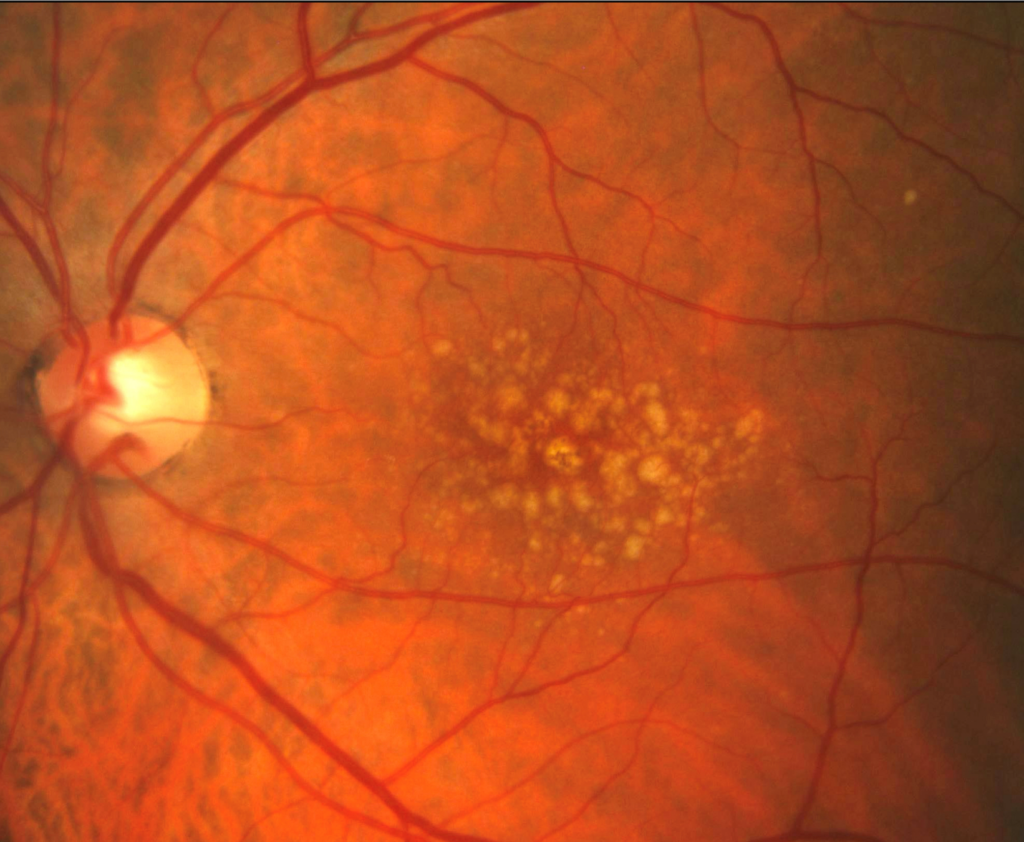
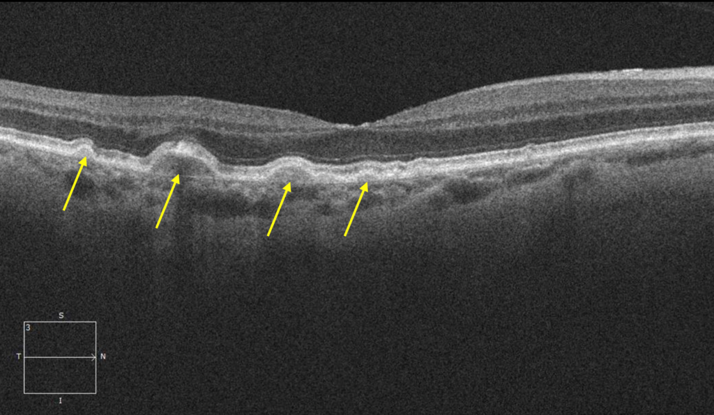
Early AMD is characterised by the presence of yellow deposits under the retina, known as drusen (Figure 1), and in some cases there is hypo- or hyper-pigmentation of the retinal pigment epithelium (RPE), the outer pigmentary layer of the retina. In intermediate AMD, larger drusen are present. Late-stage AMD is divided into the neovascular (“wet”) and atrophic (“dry”) forms.
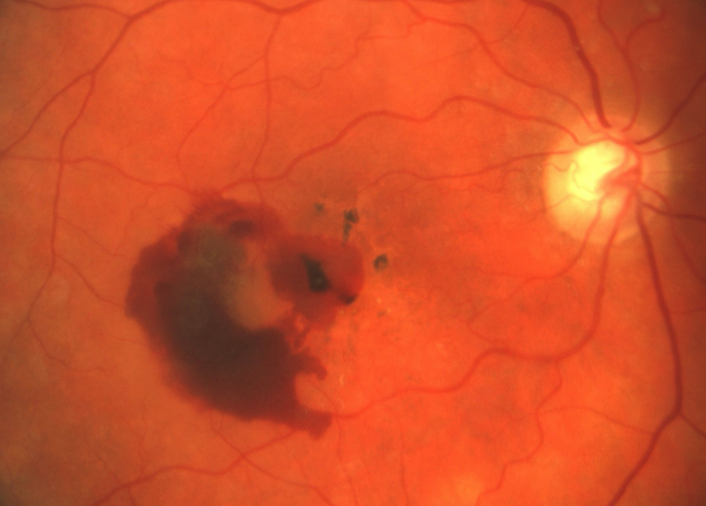
Neovascular AMD (nAMD) is characterised by growth of abnormal vessels from the choroidal (vascular) layer, extending under the layers of the retina. These can cause fluid leakage and subretinal haemorrhage (Figure 2), which can lead to permanent damage to macular photoreceptors from iron-related toxicity, impairment of diffusion of oxygen and nutriment, and mechanical damage due to clot contraction or fibrosis. Rapid central vision loss with distortion and a reduction of central vision can occur, and urgent referral to an ophthalmologist for treatment is critical.
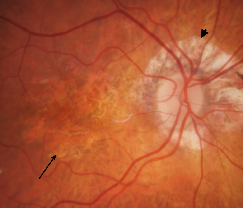
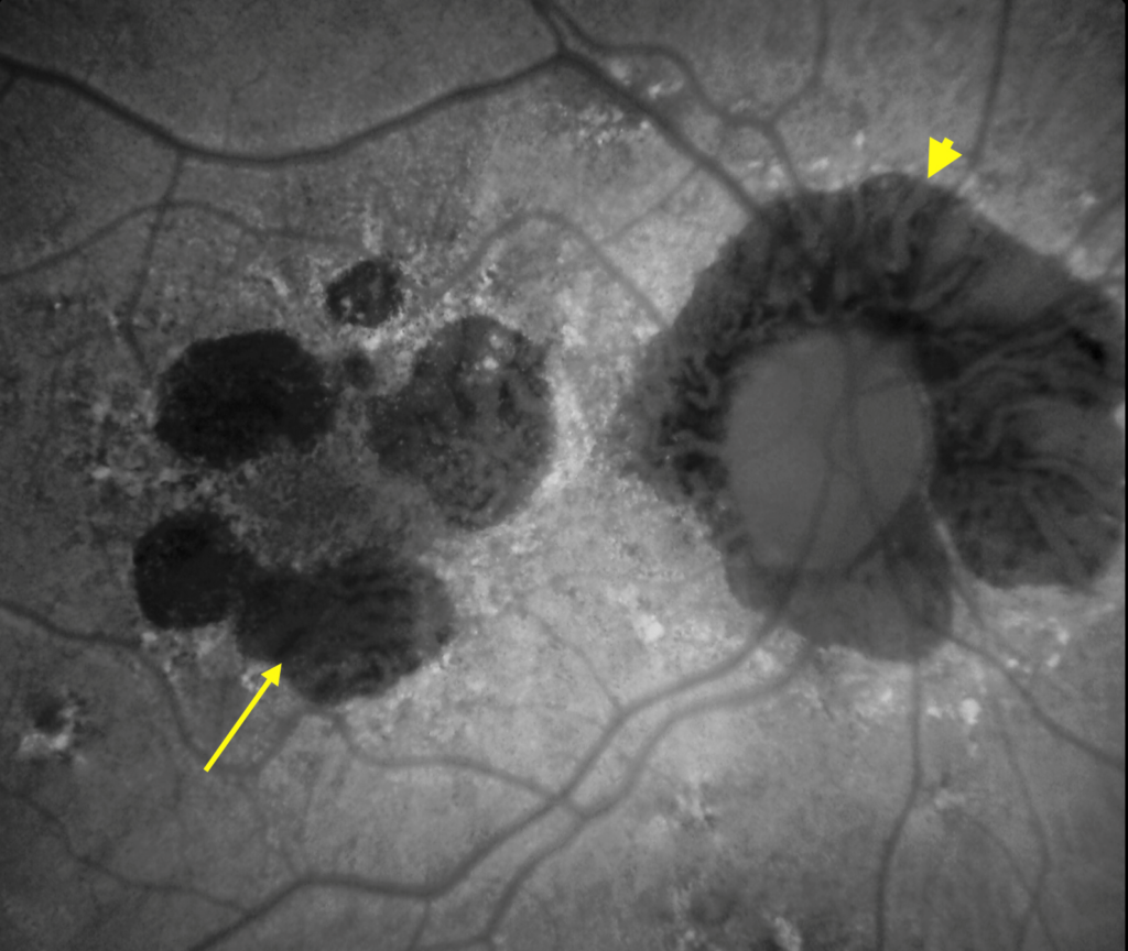
In atrophic AMD, areas of atrophy of the outer retina and RPE (“geographic atrophy” (GA)) gradually enlarge to form dull patches in the paracentral vision (Figure 3). Visual acuity usually remains unaffected until these atrophic patches extend into the centre of the macula (fovea). These changes typically develop over months to years, so any sudden vision changes are suspicious for development of neovascularisation, which can also occur in eyes with pre-existing GA.
Diagnosis and referral
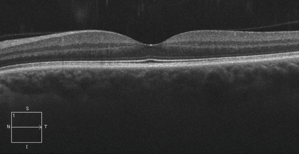
Increasingly, the diagnosis of AMD is being made in patients with few or no symptoms, by virtue of imaging studies carried out by optometrists and ophthalmologists — in particular the widespread use of optical coherence tomography (OCT), which allows for non-invasive, cross-sectional images of the macula (Figure 4).
GPs should suspect macular disease (in particular AMD) in patients complaining of central vision disturbance — in particular distortion (in straight lines or during reading), dull spots (scotomas) or difficulty reading/performing close tasks despite appropriate near correction (with spectacles). Sudden onset of these symptoms is suspicious for development of nAMD, and urgent referral is necessary. Such patients should be seen within a few days by an optometrist or ophthalmologist, as early diagnosis and treatment of nAMD is essential to obtain the best possible results from intravitreal therapy (see below).
Diagnosis is confirmed by clinical examination of the macula, OCT scanning/OCT angiography and, in selected cases, fluorescein or indocyanine green angiography.
Diet and supplements
There is no preventive treatment for AMD. The Age-Related Eye Disease Studies (AREDS) showed a modest benefit from dietary supplementation with antioxidants, zinc and the carotenoids lutein and zeaxanthin, in select groups of patients with AMD (here and here). These supplements are not of proven benefit in early AMD and should only be prescribed if a patient has had their AMD accurately staged.
A healthy balanced Mediterranean style diet rich in coloured vegetables, fruit, and fish high in omega-3 is advocated for macular health. Eating a handful of nuts per week, limiting fats and oils (and using mainly extra virgin olive oil) and limiting alcohol intake are also recommended. These dietary patterns are associated with a range of other health benefits.
Treatment
The breakthrough in AMD treatment occurred 20 years ago with the introduction of intravitreal injections of anti-vascular endothelial growth factor (anti-VEGF) medications for nAMD. These have greatly improved the prognosis for nAMD, with vision being stabilised in most patients and with some improvement, at least in the first two years, in around 30% (here and here). This is in stark contrast to the untreated history, with 75% of eyes with nAMD losing vision to <6/60 after 3 years. There are currently four anti-VEGF agents for nAMD that are approved by the Therapeutic Goods Administration and listed on the Pharmaceutical Benefits Scheme : ranibizumab, aflibercept, brolucizumab and faricimab. These are given by intravitreal injection — initially every four weeks then at treatment intervals tailored to individual treatment responses, which are highly variable.
Anti-VEGF injections suppress neovascular growth and exudation, but are not a cure, so most patients require indefinite treatment. Most patients are able to have their treatment intervals extended to eight weeks or longer, but a small number require ongoing injections at frequent intervals to maintain their vision. Some patients are fortunate enough that treatment may be suspended, but there is a high recurrence rate, and treatment will then be restarted. Vision outcomes are comparable between the available agents.
Persistence with treatment is a major problem. Twenty per cent of nAMD patients stop treatment by the one-year mark, and this increases to 50% at five years. Factors include high treatment burden (especially as second eye involvement is common), affordability and access, perceptions that treatment is not of ongoing benefit, and fear of injections/pain or discomfort from treatment. Although daunting, intravitreal injections should not be painful.
As trusted medical advisers, GPs have a crucial role in encouraging patients to persist with treatment. Support and education for patients and carers are essential, and the Macular Disease Foundation Australia (MDFA) provides advocacy and extensive resources for AMD sufferers and their families.
There is currently no available treatment for atrophic AMD in Australia. Two intravitreal agents (pegcetacoplan and avacincaptad pegol, both complement inhibitors) have been approved in the United States, and numerous other agents for GA are in clinical trials worldwide.
Research and future developments
For nAMD treatment, there is a focus on improving durability with longer-acting agents and sustained delivery systems, and better vision outcomes with new and combination therapies. Numerous agents for GA are in clinical trials, addressing the complement pathway and various other targets. Neuroprotection, RPE transplantation and subretinal gene therapy are also being investigated.
About Macula Month
Macula Month is Macular Disease Foundation Australia’s annual campaign each May to raise awareness of macular disease – Australia’s leading cause of severe vision loss and blindness.
Some useful resources: MDFA News, Check My Macula
Associate Professor Alex Hunyor is a retinal specialist and chair of the medical committee of Macular Disease Foundation Australia.
Associate Professor Anthony Kwan is a retinal specialist and chair of the research committee of Macular Disease Foundation Australia.
The statements or opinions expressed in this article reflect the views of the authors and do not necessarily represent the official policy of the AMA, the MJA or InSight+ unless so stated.
Subscribe to the free InSight+ weekly newsletter here. It is available to all readers, not just registered medical practitioners.
If you would like to submit an article for consideration, send a Word version to mjainsight-editor@ampco.com.au.

 more_vert
Thanks for this very clear and helpful summary.